
Page 58 - Cote clinical veterinary advisor dogs and cats 4th
P. 58
Abscess, Lung 5
Abscess, Lung Client Education
Sheet
VetBooks.ir Initial Database Diseases and Disorders
• Nonspecific signs of illness (anorexia, weight
BASIC INFORMATION
loss) with fever • Complete blood count: possible neutrophilic
Definition leukocytosis with or without a left shift;
A localized collection of exudate due to sup- PHYSICAL EXAM FINDINGS anemia (usually mild, nonregenerative)
puration of lung tissue, resulting in pulmonary • Poor body condition possible with chronic abscessation
cavitation • Fever may be present, but absence does not • Survey thoracic radiographs
rule out the diagnosis. ○ Mass within pulmonary parenchyma;
Synonym • Thoracic auscultation cavitation/gas in the lesion is pathogno-
Pulmonary abscess ○ ± Loud bronchovesicular sounds or crackles monic.
○ Muffled heart/lung sounds if pyothorax ○ Consolidated lung lobe may result from
Epidemiology or pneumothorax chronic abscessation.
SPECIES, AGE, SEX • Tachypnea or dyspnea ○ ± Pneumothorax
Dog and cat (more common), any age, either sex ○ Pleural effusion (if pyothorax)
Etiology and Pathophysiology • Analysis of pleural effusion, if present (pp.
GENETICS, BREED PREDISPOSITION • Foreign body migration 1164 and 1343)
May be more common in hunting dogs due • Pneumonia (primary bacterial, fungal, ○ Cytologic evaluation
to field work aspiration) ○ Aerobic and anaerobic bacterial culture
• Parasitic infestation (Paragonimus) and susceptibility testing
RISK FACTORS • Primary pulmonary neoplasia
Foreign body inhalation Advanced or Confirmatory Testing
DIAGNOSIS CT scan
GEOGRAPHY AND SEASONALITY • Assess involvement of other intrathoracic
Inhalation of plant foreign body (e.g., grass Diagnostic Overview structures; superior visualization compared
awn) in endemic area (p. 398) The diagnosis is suggested based on patient to radiographs, especially when pleural
signalment, history, and physical examination effusion is present.
ASSOCIATED DISORDERS findings. Confirmation requires 1) thoracic ○ Additional pulmonary involvement
Hypertrophic osteopathy (p. 508) radiographs to demonstrate the pulmonary mass ○ Mediastinal abscess
• Reported in dogs with chronic pulmonary and 2) ultrasound to help differentiate abscess ○ Pleural involvement
abscessation from neoplasia. Computed tomography (CT) ○ Presence of foreign material and migration
• Causes slowly progressive lameness scanning may be necessary to help confirm the to distant sites
• May be associated with pyothorax that diagnosis, determine extent of disease, and • Assess if lesion is amenable to surgical
develops secondary to the lung abscess direct surgical intervention. Foreign bodies may resection.
• May be associated with spontaneous pneu- be located in sites distant from the abscess and • Possibly identify cause of abscess.
mothorax can be diagnosed with CT. • Rule out other causes of pulmonary
mass.
Clinical Presentation Differential Diagnosis
HISTORY, CHIEF COMPLAINT Other possible masses in the pulmonary TREATMENT
• Chronic, progressive respiratory signs: cough, parenchyma:
increased respiratory effort • Neoplasia Treatment Overview
• Acute dyspnea or respiratory decompensa- • Cyst Surgical resection of the affected lung lobe(s)
tion: rupture of abscess resulting in pneu- • Granuloma and culture-directed, long-term antimicrobial
mothorax or pyothorax • Parasitic nodules therapy generally is the treatment of choice.
A B
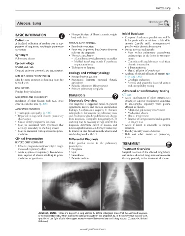
ABSCESS, LUNG Thorax of a dog with a lung abscess. A, Lateral radiograph shows that the abscessed lung area
is the right middle lobe, which overlies the cardiac silhouette in this projection. B, In the dorsoventral thoracic view,
opacities of the right middle lobe suggest extensive consolidation consistent with lung abscess. (Courtesy Dr. Richard
Walshaw.)
www.ExpertConsult.com