
Page 63 - Cote clinical veterinary advisor dogs and cats 4th
P. 63
8 Abscess, Periapical (Tooth Root)
• The periapical abscess may develop directly ○ Acute abscess (arising directly from a Drug Interactions
as a result of pulp necrosis but more com- necrotic pulp) may show only mild widen- Adverse reactions to systemic antibiotics and/
VetBooks.ir or cyst. ○ Chronic abscess is associated with bone Possible Complications
ing of apical periodontal space.
or analgesics
monly originates from a periapical granuloma
destruction (i.e., periapical lucency)
around the apex of the tooth; resorption
DIAGNOSIS
of the most apical portion of the tooth • Incomplete extraction
• Endodontic: incomplete removal of pulp,
Diagnostic Overview root may be present; the width of the substandard obturation, and restoration
An acute periapical abscess should be suspected root canals of affected teeth may be wider
when a patient presents with fever, regional compared to the root canals of unaffected Recommended Monitoring
lymphadenitis, facial swelling, or intraoral or contralateral teeth. • Physical examination
extraoral draining tracts. A chronic periapical • Radiographic evaluation to confirm healing
abscess should be suspected when a patient TREATMENT
presents with facial swelling or draining tracts. PROGNOSIS & OUTCOME
Confirmation requires meticulous oral examina- Treatment Overview
tion under general anesthesia to identify teeth The goal of treatment (extraction or endodontic Excellent if therapeutic goals (drainage
with pulpal lesions and dental radiography to treatment of the affected tooth) is to achieve and removal of cause of inflammation) are
assess the periapical status of affected teeth. drainage and remove the cause of the inflam- achieved
matory reaction.
Differential Diagnosis PEARLS & CONSIDERATIONS
• Periapical granuloma: periapical bone lysis; Acute General Treatment
accumulation of mononuclear inflammatory • Extraction or endodontic therapy (total Comments
cells, fibroblasts, and collagen pulpectomy and root canal filling) • Chronic abscess is more common than acute.
• Periapical (radicular) cyst: periapical bone • Endodontic therapy may need to be staged • An abscess associated with a draining tract
lysis; bone walls covered by cystic epithelium (i.e., performed in several sessions). usually causes less discomfort.
• Osteomyelitis: diffuse, regional bone lysis; • Concurrent systemic antibiotic treatment • Draining tracts commonly occur at the
inflammation of bone and bone marrow (e.g., 2 weeks of amoxicillin/clavulanic acid mucogingival junction on the labial or buccal
13.75 mg/kg PO q 12h or clindamycin, aspect of the tooth.
Initial Database 5.5 mg/kg PO q 12h) is indicated if fever • An acute periapical abscess with systemic
• Complete blood count, serum chemistry and/or regional lymphadenopathy exist, the signs and diffuse swelling (cellulitis) is a true
panel, urinalysis: preoperative and usually associated swelling is diffuse, or cellulitis emergency.
unremarkable (± leukocytosis) occurs. • If extraoral, the draining tract of an abscessed
• Meticulous oral examination under general • The use of systemic antibiotics alone is NOT maxillary fourth premolar tooth in a dog
anesthesia to identify teeth with pulpal appropriate treatment. usually is situated in the cheek skin at the
lesions (fractured teeth, often with pulp • Analgesic treatment with nonsteroidal level of the medial canthus of the eye, while
exposure; discolored teeth; caries; drainage antiinflammatory drugs (e.g., in dogs: the same location can be related to abscessed
tracts) (p. 1140) meloxicam 0.2 mg/kg PO q 24h; or carpro- maxillary canine or fourth premolar teeth
fen 2.2 mg/kg PO q 12h) in the cat.
Advanced or Confirmatory Testing
• Dental radiography to assess periapical status Chronic Treatment Prevention
of affected teeth (variable radiographic • Extraction or endodontic therapy • Minimize the risk of dental trauma.
appearance depending on whether acute or • Monitor teeth at risk for pulpal disease (e.g., • Monitor (clinically and radiographically)
chronic process) uncomplicated crown fractures). teeth that have been subjected to trauma
A B C
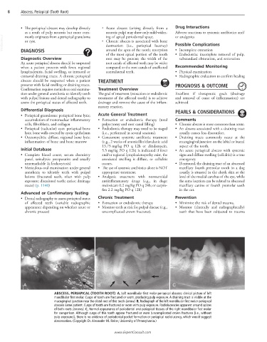
ABSCESS, PERIAPICAL (TOOTH ROOT) A, Left mandibular first molar periapical abscess: clinical picture of left
mandibular first molar. Cusps of tooth are fractured or worn, producing pulp exposure. A draining tract is visible at the
mucogingival junction near the distal root of this tooth (arrow). B, Radiograph of the left mandibular first molar periapical
abscess same patient. Cusps of tooth are fractured or worn with pulp exposure. Radiolucencies apparent around apices
of both roots (arrows). C, Normal appearance of periodontal and periapical tissues of the right mandibular first molar
for comparison. Although cusps of this tooth appear fractured or worn (uncomplicated crown fractures [i.e., without
pulp exposure]), there is no evidence of periodontal pocket formation or periapical radiolucency, which would suggest
abscessation. (Copyright Dr. Alexander M. Reiter, University of Pennsylvania.)
www.ExpertConsult.com