
Page 769 - Cote clinical veterinary advisor dogs and cats 4th
P. 769
Fractures of the Mandible and Maxilla 363
DIAGNOSIS TREATMENT ○ If the mandible is displaced, resulting
in malocclusion, treatment is required;
Treatment Overview
VetBooks.ir The diagnosis is suspected based on history Acutely, all patients should receive adequate splinting such as composite bridge between Diseases and Disorders
Diagnostic Overview
noninvasive techniques first (interarch
maxillary and mandibular canine teeth)
and physical examination; the fracture may be
analgesia (systemic and/or local). Immediate
grossly apparent or may be subtle. Radiography
application of a custom-made muzzle. Long-
under general anesthesia is often confirmatory fracture stabilization can be achieved with before considering placing intraosseous
wires or bone plates.
and is indicated in all cases to determine term goals are to provide fracture reduction ○ Condylar process fractures may require
optimal treatment. Nutritional support can be and fixation to restore dental occlusion and condylectomy if there is progressive
important if the patient cannot prehend and oral functions. difficulty in opening the mouth (tem-
swallow food, and esophagostomy tube place- poromandibular joint ankylosis).
ment may be performed under general anes- Acute General Treatment • Maxilla
thesia before or immediately after jaw fracture • Teeth may need to be removed to allow ○ Interdental wiring and intraoral composite
management. proper occlusion or closure of soft-tissue splint, intraosseous wiring, bone plating
defects. ○ Midline palatal separation: primary
Differential Diagnosis • Mandibular body (surgical) soft-tissue closure if no tension;
• Trigeminal neuritis/neuropathy/mandibular ○ Muzzling in stable or minimally displaced interquadrant splinting (wire-reinforced
neurapraxia (cranial nerve V) fracture: muzzle should be flexible (nylon composite splint) if severe distraction
• Temporomandibular joint luxation or white cotton hospital-type tape) and • Adjunctive treatment: broad-spectrum anti-
• Open-mouth jaw locking be sufficiently snug to immobilize fracture biotic therapy (e.g., amoxicillin-clavulanate
• Primary dental/periodontal disease while still allowing the patient to drink 13.75 mg/kg PO q 12h) for 1-2 weeks with
• Neoplasia water and lick liquid food. Favorable open/contaminated fractures
• Foreign body mandibular fractures in animals < 6-8
months old often do not require treatment Chronic Treatment
Initial Database other than suturing of torn soft tissues • Teeth causing mild malocclusion can be
• CBC/serum chemistry panel: generally and placing a tape muzzle for 2-3 weeks. surgically reduced (without pulp exposure)
unremarkable ○ Interdental wiring and intraoral composite or extracted.
• Head radiographs of stable, sedated patient splint (preferred noninvasive technique of • Tape muzzles and sutures through labial
can provide limited information. jaw fracture repair) buttons are removed in 2-6 weeks, composite
• Open-mouth, oblique, lateral, and ventro- ○ Intraosseous/interfragmentary wiring bridges in 5-8 weeks.
dorsal views, and intraoral dental radiographs ○ Circumferential wire for symphyseal • Partial mandibulectomy possible for chronic
are preferred, and general anesthesia is usually separation/perisymphyseal fracture nonunions
necessary. ○ External skeletal fixation • Oronasal fistulas (p. 720) may need second-
• Thoracic/abdominal radiographs to delineate ○ Bone plating ary or delayed closure.
other traumatic lesions • Mandibular ramus • Adjunctive treatment: oral instillation of
• Cranial nerve examination ○ Fractures rarely require any particular dilute (0.12%) chlorhexidine solution or
treatment beyond muzzling (snug tape gel for 2-4 weeks; brushing of teeth and
Advanced or Confirmatory Testing muzzle through which the patient can intraoral splints
CT provides high resolution for maxillary still eat/drink) or modified labial button
and caudal mandibular fractures (but a proper technique (sutures attach buttons on the Nutrition/Diet
diagnosis can be obtained without CT in most left and right upper lips to a button on Provide nutritional support during healing.
cases). the chin). Blenderize food into liquid slurry, or place
A B C D
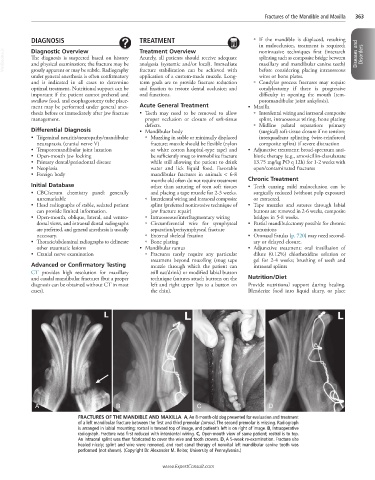
FRACTURES OF THE MANDIBLE AND MAXILLA A, An 8-month-old dog presented for evaluation and treatment
of a left mandibular fracture between the first and third premolar (arrow). The second premolar is missing. Radiograph
is arranged in labial mounting; rostral is toward top of image, and patient’s left is on right of image. B, Intraoperative
radiograph. Fracture was first reduced with interdental wiring. C, Open-mouth view of same patient; rostral is to top.
An intraoral splint was then fabricated to cover the wire and tooth crowns. D, A 5-week re-examination. Fracture site
healed nicely; splint and wire were removed, and root canal therapy of nonvital left mandibular canine tooth was
performed (not shown). (Copyright Dr. Alexander M. Reiter, University of Pennsylvania.)
www.ExpertConsult.com