
Page 764 - Cote clinical veterinary advisor dogs and cats 4th
P. 764
360 Fractures of the Femur
PHYSICAL EXAM FINDINGS • Spinal trauma: bilateral hindlimb paresis/ nail, intramedullary pins/cerclage, or plate/
rod.
• Lameness of affected limb paralysis, abnormal reflexes • Supracondylar fractures are repaired with
VetBooks.ir • Crepitation/pain at hip/stifle joint Initial Database lag screws, pins, condylar plates, or locking
• Swelling, bruising, or shortening of limb
• Craniocaudal and lateral radiographs of
plates.
• Loss of sensation to medial (femoral nerve)
affected hindlimb and pelvis ± lumbar spine
or lateral (sciatic nerve) digits due to regional
swelling, bruising, and transient peripheral • Abdominal and thoracic radiographs if • Trochanteric fractures are repaired with pin
and tension-band wiring or lag screws to
nerve dysfunction whole-body trauma counteract pull of gluteal muscles.
• There may be evidence of trauma to • Evaluation of medial/lateral sensations of • Femoral neck and capital physeal fractures
other parts of the body, or even signs of digits are repaired with multiple small pins or lag
shock. • Comprehensive neurologic exam (p. 1136) screw(s).
to evaluate for spinal trauma • Femoral head and neck excision (FHNE; also
Etiology and Pathophysiology • CBC, serum biochemistry panel, and uri- known as femoral head ostectomy [FHO])
• Most common (45%) long-bone fracture nalysis to assess anesthetic risk; see American can be performed for neck and capital physeal
• Concurrent injuries to abdominal wall Society of Anesthesiologists classification fractures in cats and small dogs.
or organs, pelvis, and lumbar spine are (p. 1196) • Total hip replacement may be considered for
common. certain fractures of the femoral head and/or
• Capital physeal fracture (immature animals) Advanced or Confirmatory Testing neck.
disrupts ascending vessels and compromises Ultrasound-guided aspirate or biopsy if
healing. pathologic fracture suspected radiographically Chronic Treatment
• Extensive hemorrhage with midshaft fractures • Restricted activity until radiographs at 4-6
contributes to shock. TREATMENT weeks to assess healing
• Radiograph early for suspected complications
DIAGNOSIS Treatment Overview based on clinical signs of recurrent lameness,
The goals of therapy are restoration of limb fever, limb swelling, peri-incisional draining
Diagnostic Overview function and alignment and reconstruction tracts
Diagnosis is based on a history of trauma and of damaged articular surfaces. Major trauma • FHNE for failed proximal repairs
on typically severe to non–weight-bearing may cause comorbid conditions that require
hindlimb lameness, proximal limb pain, soft- emergent therapy (e.g., hemorrhage, shock [p. Possible Complications
tissue swelling, and discoloration. Plain 433 and 911]) • Malunion (especially external rotation of
radiography is confirmatory. the hip with shaft fractures), nonunion
Acute General Treatment (inadequate fixation) (p. 357)
Differential Diagnosis • External coaptation (e.g., casts, splints [p. • Degenerative joint disease (articular fractures)
• Coxofemoral luxation (radiography can 1161]) is usually ineffective and carries a • Sciatic nerve damage (with retrograde
elucidate the lesion) high likelihood of inducing complications. intramedullary pin placement)
• Acetabular/pelvic fracture (radiography can • Fractures involving a joint require accurate • Decreased hip motion with femoral head
elucidate the lesion) reconstruction. and neck excision
• Bone neoplasia and pathologic fracture • Midshaft fractures are stabilized with bone • Implant failure
(radiography can elucidate the lesion) plate/screws, external fixator, interlocking • Infection
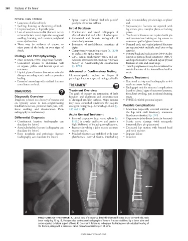
A B C
FRACTURES OF THE FEMUR A, Lateral view of transverse, distal-third femoral fracture in an 18-month-old, male
boxer weighing 20 kg. B, Postoperative ventrodorsal radiograph of femoral fracture stabilized by a bone plate and
screws applied to lateral aspect of bone. C, One-month follow-up radiograph illustrating normal endosteal healing of
the fracture, along with a periosteal callus (arrow) on medial aspect of bone.
www.ExpertConsult.com