
Page 759 - Cote clinical veterinary advisor dogs and cats 4th
P. 759
358 Fractures, Abnormal Healing
• Abnormal bone growth secondary to soft- by injection of bone marrow, autogenous skeletal fixation, and interlocking nail are
alternatives.
tissue abnormalities cancellous bone grafting, or commercially ○ Circular and linear fixators may be used by
VetBooks.ir Initial Database • Medical or nutritional disorders that could experienced surgeons to lengthen limbs or
available substrates containing bone mor-
• Osteoproliferative diseases and neoplasia
causing bone deformity
phogenetic proteins (BMPs).
transport bone across a gap by distraction
osteogenesis.
Radiographs: good-quality orthogonal views negatively impact bone healing should be • Bone grafting is critical for successful treat-
addressed.
are essential. Radiographic union may lag Treatment for a nonunion requires eliminating ment of atrophic nonunions to fill in defects,
behind clinical union, especially for fractures factors that negatively affect healing and replac- promote osteogenesis, and hasten healing time.
involving metacarpal and metatarsal bones in ing them with factors that promote healing. • Aggressive physical therapy is appropriate
both species and radii in cats. Fracture stability The goal is to correct mechanical or biological for the recovery period.
and limb function must be assessed every time inadequacies and jumpstart the process of Many malunions do not require treatment;
radiographs are taken. fracture healing. those that do are addressed with corrective
• For growing animals, radiographs are com- • Identify the reason(s) for the nonunion: osteotomy (referral to an orthopedic specialist
monly taken 3-6 weeks after fracture repair ○ Mechanical: motion at the fracture site is recommended).
and thereafter at 3-4 week intervals until ○ Biological: excessive fracture gap, necrotic
healing is documented. For most fractures, bone, infection, poor vascularity Nutrition/Diet
union is expected within 3-12 weeks after • Improve the fracture environment: A high-quality, species-specific diet should
fracture stabilization, depending on the ○ Culture for infection, and treat it if be adequate to promote bone healing. Use of
patient’s age. present. excessive supplements or high-caloric diets can
• For adult animals, radiographs are commonly ○ Remove loose/broken implants. be detrimental.
taken 6-8 weeks after fracture repair and ○ Viable nonunions: removal of excessive
thereafter at monthly intervals until healing callus is not required but may allow Possible Complications
is documented. For most fractures, union better plate contouring. Debridement of • Failure of nonunions to heal despite
is expected by 16-18 weeks after fracture fracture ends and bone grafting is usually intervention
stabilization. not necessary. • Cancellous bone graft donor site morbidity:
• With delayed/nonunion, there is lack of ○ Atrophic nonunions: debride fibrous tissue pain or (rarely) iatrogenic fracture
progression or no bony healing over a and fracture ends until some bleeding is • Implant failure, bone healing problems, or
period of months. The time to union also evident from the bone. This provides soft-tissue morbidity at corrective osteotomy
is influenced by the type of fracture fixa- mesenchymal cells and promotes ingrowth sites
tion used for stabilizing the fracture. Bones of vascular supply. It also provides better
stabilized with less rigid implants (e.g., casts, fracture apposition and allows compres- Recommended Monitoring
intramedullary pins) normally produce more sion of the fracture site. Add autogenous • Monthly follow-up radiographs until union
callus and are radiographically healed sooner cancellous bone graft or other substances is documented
than with more rigid types of fixation (e.g., to promote bone healing. • Limb use and fracture site stability should
dynamic compression plating). • Rigid fixation is essential: be assessed monthly until union occurs.
• With malunion, shortening or angulation of ○ Bone plate (DCP or locking) stabiliza- • If infection was detected, cultures should be
a bone may be evident. Torsional malunions tion is considered the fixation method of taken 6 weeks after antibiotic therapy was
cause internal or external rotation of joints. choice; plate/rod combination, external instituted.
Advanced or Confirmatory Testing
• Aerobic and anaerobic culture of the bone by
fine-needle aspiration if infection is suspected
• Scintigraphy can be used to evaluate the
fracture site for activity.
• CT or MRI (p. 1132) may aid in making
the diagnosis and in monitoring healing.
Three-dimensional (3D) reconstructive
techniques may aid in diagnosis and in
treatment planning.
TREATMENT
Treatment Overview
The goal of treatment for delayed union or
nonunion is to achieve fracture union. This
requires accurate diagnosis of the underlying
factors that resulted in impaired fracture healing.
Acute General Treatment
Treatment for a delayed union is aimed at aug-
menting or continuing the original therapeutic
plan, unless the original plan was inadequate
(e.g., external coaptation) for the fracture type. A B
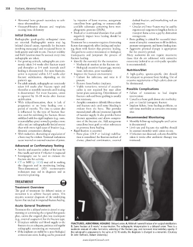
• If implant stability and blood supply to the FRACTURES, ABNORMAL HEALING Delayed union. A, Midshaft humeral fracture after surgical stabilization.
fracture site are adequate, patience and regular B, Same fracture at 8 weeks’ re-examination. The radiographic interpretation is a delayed union. There is a
radiographic monitoring are warranted. moderate amount of callus formation, widening of the fracture gap, and increased intramedullary opacity. If
• If the implants are stable but a poor biological the radiographic appearance is the same at 12-16 weeks, the diagnosis is changed to a nonunion. (Courtesy
environment exists, healing can be promoted Dr. John Hathcock, Auburn University.)
www.ExpertConsult.com