
Page 756 - Cote clinical veterinary advisor dogs and cats 4th
P. 756
356 Foreign Body, Respiratory Tract
• Cytologic evaluation/culture of nasal cavity ○ Lobectomy for lung lobe consolidation, • Pulmonary abscess or recurrent pyothorax
(by deep nasal swab or tissue biopsy) to bronchopneumonia, or bronchoesophageal if migrating FB remains or inappropriate
VetBooks.ir rhinitis. Culture of nasal secretions is not culture (aerobic and anaerobic) of excised • Chronic rhinitis possible if turbinates are
fistula; histopathologic evaluation and
antibiotics used
identify organisms associated with secondary
removed during rhinotomy
recommended.
tissue
• Nasal CT: mucosal thickening, focal bone
thickening and destruction; may not dif- • For pyothorax or bronchopneumonia, broad- • Tracheal rupture or tear
spectrum antibiotics
ferentiate FB rhinitis from nasal aspergillosis. • For pyothorax, bilateral large diameter Recommended Monitoring
Small and/or soft-tissue density FB seldom thoracostomy tubes or thoracotomy (median • Tracheal/bronchial/lung/thoracic FB: repeat
identified on CT. sternotomy for generalized disease) and radiographs or endoscopy if clinical signs
• Nasopharyngeal FB may be visible in anes- thoracic lavage/drainage (p. 857) recur.
thetized patients by soft palate retraction ○ If tubes, lavage with sterile, lukewarm • Pyothorax or pneumonia: repeat radiographs
during oropharyngeal exam (p. 1125) or isotonic fluids (20 mL/kg) q 12h for 5-7 1 week after discontinuing antibiotics or if
retroflexed nasopharyngoscopy. days clinical signs recur.
• Bronchoscopy (p. 1074) is generally diag- • Surgical exploration if intrathoracic mass,
nostic for tracheal foreign bodies and some radiographic evidence of pulmonary or medi- PROGNOSIS & OUTCOME
bronchial foreign bodies (may be obscured astinal lesions, pneumothorax, Actinomyces,
by mucopurulent exudates or beyond the or no improvement with medical therapy • Outcome is excellent if patient survives
reach of endoscope). FB extraction and secondary infections are
○ Airway lavage: neutrophilia ± bacteria on Chronic Treatment treated appropriately.
cytology for tracheobronchial FB • Antibiotics for secondary infections, based ○ Bronchopulmonary abscess develops with
○ Neutrophils are often degenerative when on culture/sensitivity FB migration if tracheobronchial grass awn
associated with bacterial infection. ○ Amoxicillin or amoxicillin plus clavulanic is present for more than 2 weeks.
acid 12.5-20 mg/kg PO q 12h for obligate • Complication and mortality rates are higher
TREATMENT anaerobes, Pasteurella spp, Actinomyces spp with chronicity.
○ Trimethoprim sulfa 10-15 mg/kg PO q • Endoscopy is successful in removing
Treatment Overview 12h (possibly higher but may increase 76%-84% of tracheobronchial FBs.
Remove FB, and treat secondary infections. risk of adverse effects), or amikacin for ○ Tracheobronchial plant material can
Animals in respiratory distress from obstruc- Nocardia spp fragment, requiring multiple endoscopic
tion may require immediate anesthesia and FB ○ For pyothorax, antibiotics are administered episodes for complete removal.
retrieval. for 1-2 months.
• Bronchodilators (theophylline, terbutaline, PEARLS & CONSIDERATIONS
Acute General Treatment or inhaled albuterol) for 3-5 days after
• Oxygen supplementation (p. 1146) if endoscopic removal of bronchial foreign Comments
needed bodies, particularly in cats. • Some nasal FBs can be removed with vigor-
• Sedation if extremely stressed (i.e., anxiety ous flushing.
is contributing to dyspnea) Possible Complications • Right bronchial systems are more likely to
○ Acepromazine (0.1-0.5 mg total dose • Inability to oxygenate during endoscopy or be affected by inhaled bronchial FBs because
IM or IV) or dexmedetomidine if not surgery of direct tracheobronchial path; however,
systemically ill • Worsening of obstruction with flushing, 21% of affected dogs have more than one
○ Butorphanol 0.2-0.4 mg/kg IV may be FB manipulation, or endoscopic trauma tracheobronchial FB.
given additionally q 2-4h as needed (mucosal swelling) • If sulfur granules or branching, filamentous,
• Rehydrate as needed. • Noncardiogenic pulmonary edema may gram-positive rods suggestive of Actino-
• Thoracocentesis if respiratory compromise develop within hours after FB removal. myces infection are seen during cytologic
from pleural effusion or pneumothorax
(p. 1164)
• Immediate anesthesia and FB removal if
trachea severely obstructed
○ Distal tracheostomy (p. 1166) may be
needed for short-term airway management.
• If possible, nonsurgical (often endoscopic)
removal of nasal, laryngeal, tracheal, and
some bronchial FBs
○ Nasal FB sometimes removed during
retrograde or antegrade lavage/flush
○ Tracheal FB in cats removed with
custom-made, 24-inch grasping forceps
under fluoroscopic guidance. Successful
retrieval with tracheobronchoscopy and
alligator forceps through scope channel
in 83% of cats
○ Some tracheal FBs removed with vacuum
suction or passage of balloon (Fogarty) A B
catheter beyond the FB and then inflation
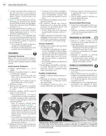
and retraction of balloon FOREIGN BODY, RESPIRATORY TRACT CT scan of a 3-year-old, intact, male Siberian Husky–cross dog
• Rhinotomy (dorsal or ventral), tracheotomy, with chronic cough. On axial (A) and sagittal reconstruction (B), there is a suggestion of an intrabronchial
bronchotomy, or lung lobectomy for non- foreign body (arrows). There is substantial peribronchiolar and alveolar infiltrate. A large piece of wheat was
retrievable intraluminal FB removed bronchoscopically.
www.ExpertConsult.com