
Page 751 - Cote clinical veterinary advisor dogs and cats 4th
P. 751
354 Foreign Body, Linear Gastrointestinal
contrast series because up to 16% of cats
and 41% of dogs may have perforation of
VetBooks.ir ○ The advantage of iodine (resorbed from
the intestinal tract.
the peritoneal cavity if perforation exists,
unlike barium) must be weighed against
its reduced degree of contrast, foul taste
(compliance), and the access to any
spilled barium at the time of laparotomy
because surgical intervention is indicated
if perforation is confirmed.
TREATMENT
Treatment Overview
Rehydration and rapid surgical intervention
are recommended. Specific treatment goals:
• Correct dehydration and electrolyte
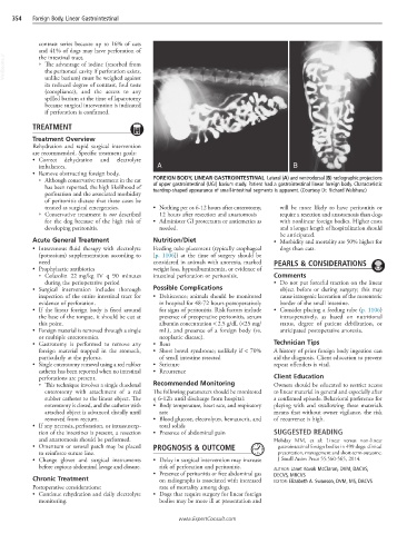
imbalances. A B
• Remove obstructing foreign body.
○ Although conservative treatment in the cat FOREIGN BODY, LINEAR GASTROINTESTINAL Lateral (A) and ventrodorsal (B) radiographic projections
has been reported, the high likelihood of of upper gastrointestinal (UGI) barium study. Patient had a gastrointestinal linear foreign body. Characteristic
teardrop-shaped appearance of small-intestinal segments is apparent. (Courtesy Dr. Richard Walshaw.)
perforation and the associated morbidity
of peritonitis dictate that these cases be
treated as surgical emergencies. • Nothing per os 6-12 hours after enterotomy, will be more likely to have peritonitis or
○ Conservative treatment is not described 12 hours after resection and anastomosis require a resection and anastomosis than dogs
for the dog because of the high risk of • Administer GI protectants or antiemetics as with nonlinear foreign bodies. Higher costs
developing peritonitis. needed. and a longer length of hospitalization should
be anticipated.
Acute General Treatment Nutrition/Diet • Morbidity and mortality are 50% higher for
• Intravenous fluid therapy with electrolyte Feeding tube placement (typically esophageal dogs than cats.
(potassium) supplementation according to [p. 1106]) at the time of surgery should be
need considered in animals with anorexia, marked PEARLS & CONSIDERATIONS
• Prophylactic antibiotics weight loss, hypoalbuminemia, or evidence of
○ Cefazolin 22 mg/kg IV q 90 minutes intestinal perforation or peritonitis. Comments
during the perioperative period • Do not put forceful traction on the linear
• Surgical intervention includes thorough Possible Complications object before or during surgery; this may
inspection of the entire intestinal tract for • Dehiscence; animals should be monitored cause iatrogenic laceration of the mesenteric
evidence of perforation. in hospital for 48-72 hours postoperatively border of the small intestine.
• If the linear foreign body is fixed around for signs of peritonitis. Risk factors include • Consider placing a feeding tube (p. 1106)
the base of the tongue, it should be cut at presence of preoperative peritonitis, serum intraoperatively, as based on nutritional
this point. albumin concentration < 2.5 g/dL (<25 mg/ status, degree of patient debilitation, or
• Foreign material is removed through a single mL), and presence of a foreign body (vs. anticipated postoperative anorexia.
or multiple enterotomies. neoplastic disease).
• Gastrotomy is performed to remove any • Ileus Technician Tips
foreign material trapped in the stomach, • Short bowel syndrome; unlikely if < 70% A history of prior foreign body ingestion can
particularly at the pylorus. of small intestine resected aid the diagnosis. Client education to prevent
• Single enterotomy removal using a red rubber • Stricture repeat offenders is vital.
catheter has been reported when no intestinal • Recurrence
perforations are present. Client Education
○ This technique involves a single duodenal Recommended Monitoring Owners should be educated to restrict access
enterotomy with attachment of a red The following parameters should be monitored to linear material in general and especially after
rubber catheter to the linear object. The q 6-12h until discharge from hospital: a confirmed episode. Behavioral preference for
enterotomy is closed, and the catheter with • Body temperature, heart rate, and respiratory playing with and swallowing these materials
attached object is advanced distally until rate means that without owner vigilance, the risk
removed from rectum. • Blood glucose, electrolytes, hematocrit, and of recurrence is high.
• If any necrosis, perforation, or intussuscep- total solids
tion of the intestines is present, a resection • Presence of abdominal pain SUGGESTED READING
and anastomosis should be performed. Hobday MM, et al: Linear versus non-linear
• Omentum or serosal patch may be placed PROGNOSIS & OUTCOME gastrointestinal foreign bodies in 499 dogs: clinical
to reinforce suture line. presentation, management and short-term outcome.
• Change gloves and surgical instruments • Delay in surgical intervention may increase J Small Anim Pract 55:560-565, 2014.
before copious abdominal lavage and closure. risk of perforation and peritonitis. AUTHOR: Janet Kovak McClaran, DVM, DACVS,
• Presence of peritonitis or free abdominal gas DECVS, MRCVS
Chronic Treatment on radiographs is associated with increased EDITOR: Elizabeth A. Swanson, DVM, MS, DACVS
Postoperative considerations: rate of mortality among dogs.
• Continue rehydration and daily electrolyte • Dogs that require surgery for linear foreign
monitoring. bodies may be more ill at presentation and
www.ExpertConsult.com