
Page 748 - Cote clinical veterinary advisor dogs and cats 4th
P. 748
352 Foreign Body, Esophageal
• Rarely, pressure of foreign object on trachea • Contrast esophageal radiographs esophagitis (p. 312). Do not give antacids
causes coughing or choking. ○ Rarely needed and may obscure visu- if bone has been pushed into the stomach
VetBooks.ir PHYSICAL EXAM FINDINGS typically better to do esophagoscopy if • Appropriate treatment of aspiration pneu-
because gastric acid is needed to dissolve
alization during esophagoscopy; it is
the bone.
plain radiographs are not diagnostic but
• Ptyalism occasionally noticed
foreign body is strongly suspected.
• Dysphagia or gagging seen rarely
• Dyspnea/fever sometimes seen secondary to ○ Risk of aspiration of contrast material or monia, pleuritis/mediastinitis, if present
perforation, mediastinitis/pleuritis, and/or leakage of contrast into the mediastinum: Chronic Treatment
aspiration pneumonia use iodide-based contrast instead of barium Mechanical dilation of strictures, if necessary
• Rarely, can palpate the foreign object in the (p. 310)
cervical esophagus Advanced or Confirmatory Testing
Esophagoscopy (p. 1098): always provides a Nutrition/Diet
Etiology and Pathophysiology definitive diagnosis Place gastrostomy feeding tube (p. 1109)
• Bones are the most common foreign body if esophagitis is so severe that the patient
in dogs. TREATMENT cannot or will not eat.
• Other common foreign objects include food,
fishhooks, rawhide treats, and dental chew Treatment Overview Possible Complications
toys. Remove the foreign object and resolve com- • Stricture (cicatrix) causing partial or complete
• Hairballs are important in cats. plications (e.g., esophagitis or perforation with esophageal obstruction
resulting pleural/mediastinal sepsis). • Esophageal perforation causing septic
DIAGNOSIS pleuritis/mediastinitis (p. 857) or uncom-
Acute General Treatment monly bronchoesophageal fistula
Diagnostic Overview • Esophageal foreign objects should be removed • Insufflation of air during esophagoscopy
History is important; ask whether the patient as soon as the patient is an acceptable can cause tension pneumothorax if there is
swallowed anything immediately before clinical anesthetic candidate to reduce chance of a perforation and/or gastric dilation if the
signs began or if it frequently chews or esophageal perforation. scope cannot be advanced into the stomach
mouths objects (i.e. has a propensity to foreign • Esophagoscopy to remove foreign object and to remove air.
body ingestion). Plain cervical and thoracic determine degree of esophagitis (p. 1098) • Severe bleeding is uncommon (and rarely life-
radiographs are the tests of choice and are ○ If foreign object cannot be removed, threatening) but possible when manipulating/
confirmatory in almost all cases (assuming attempt to push into the stomach for removing foreign object.
good radiographic technique); esophagoscopy is surgical removal or dissolution (bone).
always confirmatory and is usually therapeutic. • A Foley catheter can be used for objects Recommended Monitoring
without sharp edges (place balloon behind Be sure patient is able to eat without regurgita-
Differential Diagnosis foreign object, inflate balloon, then pull the tion within 1-2 days of foreign body removal. If
Regurgitation: catheter out so that the balloon draws the regurgitation occurs after several days, evaluate
• Esophagitis foreign object orad and out). A lubricated for possible stricture.
• Megaesophagus (acquired [idiopathic or Foley catheter can be used to open lower
secondary to systemic disease] or congenital) esophageal sphincter when pushing object PROGNOSIS & OUTCOME
• Esophageal mass into stomach.
• Esophageal stricture • Surgery if esophagoscopy is unsuccessful at • Good if there is no perforation and esopha-
• Vascular ring anomaly removing the foreign object gitis is not severe
• Prokinetics (e.g., metoclopramide 0.2- • Good to guarded if severe, near-circumferential
Initial Database 0.4 mg/kg IM, SQ, or PO q 8h) and/or esophagitis is likely to cause stricture
• CBC and serum biochemistry panel to antacid (omeprazole 1-2 mg/kg PO q 12h • Guarded to poor if severe septic mediastinitis
prepare for anesthesia or pantoprazole 1 mg/kg IV q 12h) to treat or pleuritis from perforation
• Plain thoracic radiographs
○ Differentiate esophageal foreign body
from megaesophagus (often generalized
dilation as opposed to esophageal dilation
associated with foreign body, which can
be seen cranial to the foreign body) and
other thoracic masses.
○ Almost all foreign objects can be seen with
good-quality films, but radiolucent objects
(e.g., poultry bones, rawhide) may require
excellent technique. Three views may be
needed to see some foreign objects.
○ Concurrent pneumothorax, pneumome-
diastinum, or pleural effusion is suggestive
of esophageal perforation.
○ Esophageal foreign objects can radiographi-
cally resemble pulmonary or mediastinal
lesions.
○ Assess for complications: aspiration
pneumonia and/or evidence of perforation
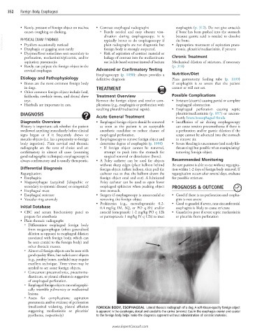
(mediastinal widening, pleural effusion FOREIGN BODY, ESOPHAGEAL Lateral thoracic radiograph of a dog. A soft-tissue-opacity foreign object
suggesting mediastinitis or pleuritis/ is apparent in the esophagus, dorsal and caudal to the carina (arrows). Gas in the esophagus cranial and caudal
pyothorax, respectively) to the foreign body helps make the diagnosis apparent without administration of contrast material.
www.ExpertConsult.com