
Page 788 - Cote clinical veterinary advisor dogs and cats 4th
P. 788
374 Gallbladder Mucocele
Video
Gallbladder Mucocele Available Client Education
Sheet
VetBooks.ir
DIAGNOSIS
• Urinalysis: generally unremarkable; biliru-
BASIC INFORMATION
binuria possible (nonspecific)
Definition Diagnostic Overview • Abdominal radiography: generally unremark-
An accumulation of bile-laden mucus within • GB mucocele can be an incidental finding able. In cases with GB rupture, decreased
the gallbladder (GB). Histologic exam of the or responsible for the presenting complaint. serosal detail may be noted.
GB wall shows cystic mucinous hyperplasia. • Abdominal US is the standard diagnostic • Abdominal US: characteristic stellate/striated
GB mucocele is the most common reason for tool. GB contents in nonruptured cases are intraluminal GB contents (see Video); subjec-
biliary surgery in dogs. hyperechoic and non–gravity dependent, tive GB distention can be noted. The GB
with a classic stellate pattern (hence the wall should be assessed for integrity.
Synonyms terms kiwi or starfish GB). • After GB rupture, free abdominal fluid may
Kiwi gallbladder, cystic mucinous hyperplasia • GB rupture may be associated with focal be seen; the fluid should be analyzed for
of the gallbladder or generalized peritonitis, indicated by evidence of sepsis and/or bile peritonitis.
fluid around the GB (+/− elsewhere in the Concurrent pancreatitis and inflammation
Epidemiology abdomen) and inflammation of adjacent of adjacent structures may be noted. In
SPECIES, AGE, SEX structures. rare cases, the mucocele may be free in the
Older, small or medium-sized dogs; no abdominal cavity.
sex predisposition reported; one feline case Differential Diagnosis
reported For vomiting with elevated liver enzyme activi- Advanced or Confirmatory Testing
ties +/− icterus: • At surgery, GB contents are solid and rubbery
GENETICS, BREED PREDISPOSITION • Pancreatitis or semiliquid, with lumps of organized
Cocker spaniels, Shetland sheepdogs, border ter- • Acute hepatotoxicosis mucus.
riers, and miniature schnauzers are predisposed. • Cholecystitis (acute, chronic, or necrotizing) • Histopathologic exam of the GB wall
An insertion mutation in the ABCB4 gene was • Cholelithiasis confirms cystic mucinous hyperplasia.
suggested as an underlying cause, but it has not • Acute or chronic hepatitis
been verified by additional studies. For hyperechoic GB contents on US: TREATMENT
• GB sludge (gravity dependent, unlike GB
RISK FACTORS mucocele) Treatment Overview
Hyperadrenocorticism (spontaneous or iatro- • Surgical intervention is indicated in sick dogs,
genic), hypothyroidism, imidacloprid use Initial Database with cholecystectomy generally considered
• CBC: neutrophilic leukocytosis with mono- the treatment of choice.
ASSOCIATED DISORDERS cytosis if clinical signs present • Prophylactic laparoscopic or open surgical
Bile peritonitis with secondary septic peritonitis, • Serum biochemistry profile: elevated alanine cholecystectomy may be considered in stable
extrahepatic biliary obstruction aminotransferase (ALT), alkaline phosphatase patients without evidence of GB rupture or
(ALP), aspartate aminotransferase (AST), and peritonitis.
Clinical Presentation gamma-glutamyltransferase (GGT) activities. • Medical therapy can be considered in patients
HISTORY, CHIEF COMPLAINT Serum bilirubin concentration is high in most with minimal clinical compromise and no
• May be an incidental finding on abdominal clinically ill patients. Hypercholesterolemia, evidence of GB wall compromise.
ultrasonography (US) hyperglobulinemia, and hypoalbuminemia • Dogs should be screened for underlying
• Clinically affected patients often vomit, along may be noted. endocrinopathy and treated as indicated.
with inappetence, and lethargy
• Other complaints include weight loss, diar-
rhea, ptyalism, and polyuria/polydipsia.
PHYSICAL EXAM FINDINGS
Abdominal pain, ascites, tachypnea, tachycardia,
fever, and icterus are all possible in advanced
cases.
Etiology and Pathophysiology
• Likely triggered by production of abnormal
gel-forming mucins by the GB epithelium;
this changes the physical and functional
properties of GB mucus.
• Cystic mucinous hyperplasia of GB epithe-
lium is probably a response to abnormal
mucus production.
• Abnormal mucins result in accumulation of
mucus and inspissated bile.
• The association between biliary sludge and GB
mucocele formation is unclear.
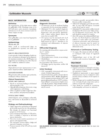
• Pressure necrosis of the GB wall may result GALLBLADDER MUCOCELE Ultrasonographic image of a gallbladder mucocele. Note the classic kiwi
in rupture. appearance.
www.ExpertConsult.com