
Page 819 - Cote clinical veterinary advisor dogs and cats 4th
P. 819
388 Glaucoma
ASSOCIATED DISORDERS reducing retinal, choroidal, and optic nerve • Ocular ultrasound if ocular media are opaque
Secondary glaucoma may follow uveitis head blood flow and axoplasmic flow in the and prevent evaluation of deeper ocular
VetBooks.ir intraocular neoplasia. • Onset and severity of these glaucomatous abnormalities (i.e., lens luxation, lens capsule
optic nerve head.
structures; helps rule out concurrent ocular
(intraocular inflammation), lens luxation, or
rupture, retinal detachment, intraocular
changes appear to be influenced by the
Clinical Presentation
duration and extent of the IOP elevation.
DISEASE FORMS/SUBTYPES • About 50% of first eyes of dogs presented masses)
Glaucoma may be classified by with the primary glaucomas are blind at TREATMENT
• Cause: primary, secondary, or congenital initial ophthalmic exam.
• Duration: acute (vision potential) versus Treatment Overview
chronic (typically blind, buphthalmic eye) DIAGNOSIS • In acute cases, it is critical that the IOP be
lowered as soon as possible to maintain or
HISTORY, CHIEF COMPLAINT Diagnostic Overview restore vision. If tonometry is unavailable,
Transient or constant red or cloudy eye with A diagnosis of glaucoma should be considered for glaucoma suspects should be referred to a
any or all of the following: any case that presents for evaluation of red eye(s), veterinary ophthalmologist (as an emergency)
• Various evidence of pain (usually present particularly if the ocular hyperemia is accompanied for IOP measurement.
when IOP > 40 mm Hg) manifesting as by mydriasis and corneal edema and especially if • Early surgical intervention may permit better
unwillingness to be handled around the face it occurs in a predisposed breed. Elevated IOP on and longer control of IOP; the clinician is
or head or blepharospasm tonometric evaluation is confirmatory. urged to consider early referral, especially
• Bumping into objects or other manifestations when vision in one eye has already been lost.
of visual impairment or blindness Differential Diagnosis • Long-term treatment of glaucoma can be
• Increased reflection from eye (i.e., tapetal Glaucoma must be differentiated from other a frustrating endeavor because the disease
reflection from dilated pupil) causes of red eye (p. 870). tends to progress despite medical therapy.
• Enlargement of the globe (buphthalmos) Diligent monitoring, regular reassessment
Initial Database of therapeutic success, and client education
PHYSICAL EXAM FINDINGS Complete ophthalmic exam (p. 1137): are critical.
Unilateral or bilateral ocular changes; initially • Tonometry (measurement of IOP): most • Therapeutic goals are to lower IOP of affected
unilateral with primary glaucoma: important diagnostic test eye to maintain vision for as long as possible,
• Corneal edema (typically diffuse) ○ Applanation tonometry (e.g., Tono-Pen) eliminate ocular pain, and treat contralateral
• Episcleral injection (dilated and tortuous ■ Rebound tonometry (e.g., TonoVet) eye prophylactically with IOP-lowering
episcleral vessels) ■ Schiøtz indentation tonometry (requires drugs to delay the median time to onset of
• Dilated pupil and sluggish to absent pupillary conversion of value from instrument to glaucoma from ≈6 months (untreated) to
light reflexes mm Hg using human calibration chart 30 months (with prophylactic treatment)
• Possible lens subluxation/luxation (look for accompanying instrument or dog/cat (primary glaucoma).
aphakic crescent) calibration chart) • A step-by-step approach to treatment is
• Variable optic nerve cupping and retinal • Normal IOP values range from 15-25 mm shown on p. 1419.
degeneration Hg; > 25 mm Hg = glaucoma (dogs and
• Corneal stria (breaks in Descemet’s mem- cats). Acute General Treatment
brane appearing as white lines within cornea; Medical:
uncommon) Advanced or Confirmatory Testing • Regardless of the type of glaucoma, the
• Referral to veterinary ophthalmologist for following may be administered initially and
Etiology and Pathophysiology ○ Additional or multiple tonometric then long term if indicated:
• Impediment to aqueous humor outflow, measurements ○ Topical beta-adrenergic antagonists (0.5%
causing elevated IOP ○ Gonioscopy (direct observation of the timolol or 0.5% betaxolol, usually q
• Elevated IOP can damage the retina (retinal iridocorneal angle) 8-12h; decreases pressure about 5-7 mm
ganglion cells) and optic nerve head by ○ Ophthalmoscopy of the ocular fundus Hg), and
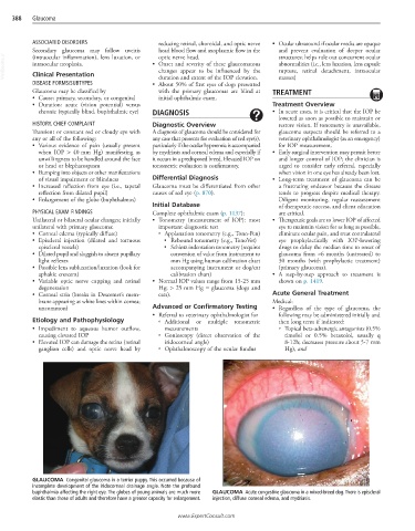
GLAUCOMA Congenital glaucoma in a terrier puppy. This occurred because of
incomplete development of the iridocorneal drainage angle. Note the profound
buphthalmia affecting the right eye. The globes of young animals are much more GLAUCOMA Acute congestive glaucoma in a mixed-breed dog. There is episcleral
elastic than those of adults and therefore have a greater capacity for enlargement. injection, diffuse corneal edema, and mydriasis.
www.ExpertConsult.com