
Page 814 - Cote clinical veterinary advisor dogs and cats 4th
P. 814
Gastrointestinal Obstruction 385
• Limited efficacy, largely replaced by Biopsy any abnormal-looking organ. Future albumin concentration < 2.5 g/dL, and
ultrasound-guided centesis course of illness may evolve in such a way presence of a foreign body (vs. neoplastic
VetBooks.ir gastric foreign body can be diagnostic and allow Chronic Treatment • Ileus Diseases and Disorders
as to make these specimens essential for
Upper GI endoscopy (p. 1098) for suspected
disease).
diagnosis.
• Short-bowel syndrome: unlikely if less than
retrieval of foreign material.
70% of small intestine resected
TREATMENT Postoperative considerations: • Stricture
• Continue rehydration and daily electrolyte • Recurrence
Treatment Overview monitoring. Treat accordingly (especially
Rehydration and rapid surgical intervention are hypokalemia arising from dilution [IV fluids] Recommended Monitoring
recommended in any patient with intestinal and anorexia). • The following parameters should be moni-
obstruction, and endoscopy can be used in • With proper technique and no contamina- tored daily until discharge from hospital:
place of surgery for some gastric obstructions. tion in straightforward cases, postoperative body temperature, blood glucose, electrolytes,
Correct dehydration and electrolyte abnor- antibiotics are not necessary and may mask abdominal pain
malities with intravenous fluid administration. signs of dehiscence and peritonitis. • The patient should be eating and not
Exploratory laparotomy is indicated to relieve • Nothing by mouth (NPO) 6-12 hours after vomiting. If anorexia or vomiting persists
the obstruction. enterotomy, 12 hours after resection and after surgery, consider performing abdomi-
anastomosis nocentesis and CBC to evaluate for possible
Acute General Treatment • Administer GI protectants as needed: development of peritonitis.
• Exploratory laparotomy for foreign body pantoprazole 0.7 mg/kg IV q 24h
retrieval or mass resection • Administer antiemetics as needed (contra- PROGNOSIS & OUTCOME
○ Some gastric foreign bodies can be indicated before resolution of obstruction).
removed by endoscopy (p. 1098). ○ Maropitant 1 mg/kg SQ q 24h • A good prognosis may be expected for
• An enterotomy may suffice for acute ○ Dolasetron 0.5 mg/kg IV, SQ q 24h acute disease, and animals with preoperative
foreign body removal; if intestinal viability ○ Metoclopramide 0.2-0.4 mg/kg PO, SQ, debilitation or shock should be given a more
is questionable, resection and anastomosis or IM q 6h (antiemetic and prokinetic) guarded prognosis.
are warranted. • Prognosis for neoplastic disease depends on
• Wide-margin (4-8 cm) resection and anas- Nutrition/Diet histopathologic grade, evidence of metastasis,
tomosis should be performed if neoplastic Feeding tube placement (p. 1106) at the time and completeness of surgical excision.
disease is suspected based on the gross of surgery should be considered in animals Lymphoma (p. 604) and adenocarcinoma
appearance of the lesion. In this case, lymph with marked weight loss, hypoalbuminemia, (p. 30) are the most frequently encountered
node and liver biopsies also are indicated. or evidence of peritonitis. If anorexia persists intestinal tumors.
• Omentum or serosal patch may be placed postoperatively, syringe feeding, feeding
to reinforce suture line. tubes, or total parenteral nutrition may be PEARLS & CONSIDERATIONS
• Change gloves and surgical instruments considered.
before abdominal lavage and closure to Comments
minimize contamination. Possible Complications Consider placing an intraoperative feeding tube
• Administer prophylactic antibiotics (cefazolin • Dehiscence: highest risk at 3-5 days postop- (esophagostomy, gastrostomy, or gastrojejunos-
22 mg/kg IV q 90 minutes during the eratively. Animals should be monitored in tomy tube [p. 1106]) in these patients based
perioperative period). hospital for 48-72 hours in the postoperative on preoperative nutritional status, degree of
• Obtain gastric, small-intestinal, and liver period for signs of peritonitis. Risk factors patient debilitation, or anticipated postoperative
biopsies, even if all appear grossly normal. include preoperative peritonitis, serum anorexia.
A B
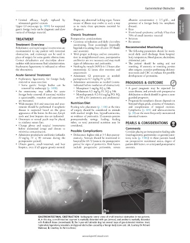
GASTROINTESTINAL OBSTRUCTION Radiographic lateral views of small-intestinal obstruction in two patients.
A, In this dog, a small-intestinal segment is markedly distended with gas (arrows), and another is markedly distended
with fluid/soft tissue (arrowheads). B, The 5-year-old Labrador dog has several loops of gas-distended intestine. Inset,
Exploratory laparotomy revealed a mid-jejunal obstruction caused by a foreign body (corn cob). (A, Courtesy Dr. Richard
Walshaw; B, Courtesy Dr. Patricia Rose.)
www.ExpertConsult.com