
Page 101 - A Practical Guide to Equine Radiography
P. 101
82 A PRACTICAL GUIDE TO EQUINE RADIOGRAPHY
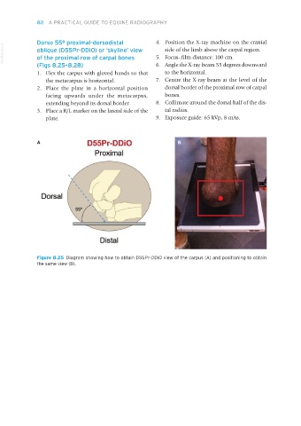
Dorso 55º proximal-dorsodistal 4. Position the X-ray machine on the cranial
VetBooks.ir oblique (D55Pr-DDiO) or ‘skyline’ view 5. Focus–film distance: 100 cm.
side of the limb above the carpal region.
of the proximal row of carpal bones
(Figs 8.25–8.28) 6. Angle the X-ray beam 55 degrees downward
1. Flex the carpus with gloved hands so that to the horizontal.
the metacarpus is horizontal. 7. Centre the X-ray beam at the level of the
2. Place the plate in a horizontal position dorsal border of the proximal row of carpal
facing upwards under the metacarpus, bones.
extending beyond its dorsal border. 8. Collimate around the dorsal half of the dis-
3. Place a R/L marker on the lateral side of the tal radius.
plate. 9. Exposure guide: 65 kVp, 8 mAs.
A B
Figure 8.25 Diagram showing how to obtain D55Pr-DDiO view of the carpus (A) and positioning to obtain
the same view (B).
Equine Radiography.indb 82 27/11/2018 11:08