
Page 145 - A Practical Guide to Equine Radiography
P. 145
126 A PRACTICAL GUIDE TO EQUINE RADIOGRAPHY
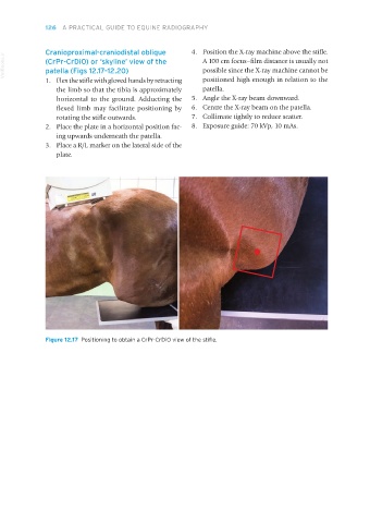
Cranioproximal-craniodistal oblique 4. Position the X-ray machine above the stifle.
VetBooks.ir (CrPr-CrDiO) or ‘skyline’ view of the A 100 cm focus–film distance is usually not
possible since the X-ray machine cannot be
patella (Figs 12.17–12.20)
1. Flex the stifle with gloved hands by retracting positioned high enough in relation to the
the limb so that the tibia is approximately patella.
horizontal to the ground. Adducting the 5. Angle the X-ray beam downward.
flexed limb may facilitate positioning by 6. Centre the X-ray beam on the patella.
rotating the stifle outwards. 7. Collimate tightly to reduce scatter.
2. Place the plate in a horizontal position fac- 8. Exposure guide: 70 kVp, 10 mAs.
ing upwards underneath the patella.
3. Place a R/L marker on the lateral side of the
plate.
Figure 12.17 Positioning to obtain a CrPr-CrDiO view of the stifle.
Equine Radiography.indb 126 27/11/2018 11:11