
Page 149 - A Practical Guide to Equine Radiography
P. 149
130 A PRACTICAL GUIDE TO EQUINE RADIOGRAPHY
Ventrodorsal under general anaesthesia − Midline views; centred on the respective
VetBooks.ir (VD GA) (Figs 13.1–13.4) anatomical landmarks:
1. Position the horse in dorsal recumbency in
a frog-leg position. ○ Tubera ischii
2. Place the plate facing upwards in a tunnel ○ Coxofemoral joints and obturadora
block under the horse’s pelvis. foramina
3. Indicate right/left with a marker. ○ Sacroiliac and lumbosacral joints.
4. Position the X-ray machine dorsal to the
pelvis. − Tuber coxae
5. Focus–film distance: 120 cm. Adjust the − Coxofemoral joints: the limb to be radio-
focus–film distance to the distance speci- graphed is tilted nearer to the plate by
fied for the grid. slightly rolling the horse.
6. Use a vertical X-ray beam.
7. X-ray beam centring depends on the area 8. Exposure guide: 150 kVp, 250 mAs.
of interest, as several overlapping views
are required for a comprehensive radio-
graphic examination of the pelvis. In a
standard adult horse, seven separate views
are described:
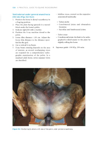
Figure 13.1 Positioning to obtain a VD view of the pelvis under general anaesthesia.
Equine Radiography.indb 130 27/11/2018 11:11