
Page 66 - A Practical Guide to Equine Radiography
P. 66
FETLOCk 47
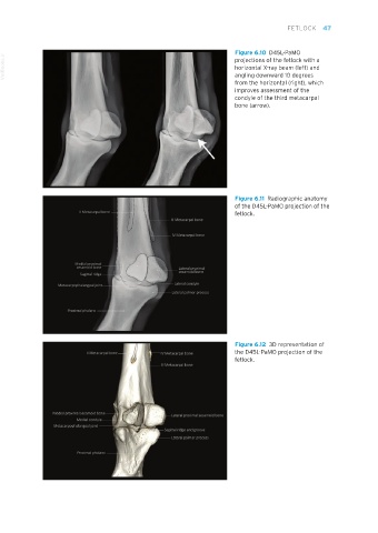
Figure 6.10 D45L-PaMO
VetBooks.ir projections of the fetlock with a
horizontal X-ray beam (left) and
angling downward 10 degrees
from the horizontal (right), which
improves assessment of the
condyle of the third metacarpal
bone (arrow).
Figure 6.11 Radiographic anatomy
of the D45L-PaMO projection of the
fetlock.
Figure 6.12 3D representation of
the D45L-PaMO projection of the
fetlock.
Equine Radiography.indb 47 27/11/2018 11:06