
Page 1125 - Small Animal Internal Medicine, 6th Edition
P. 1125
CHAPTER 62 Seizures and Other Paroxysmal Events 1097
VetBooks.ir
A B
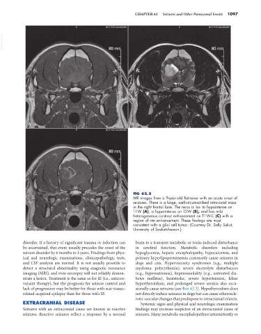
FIG 62.2
MR images from a 9-year-old Retriever with an acute onset of
seizures. There is a large, well-circumscribed intra-axial mass
in the right frontal lone. The mass is iso- to hypointense on
T1W (A), is hyperintense on T2W (B), and has mild
heterogeneous contrast enhancement on T1W-C (C) with a
region of rim enhancement. These findings are most
C consistent with a glial cell tumor. (Courtesy Dr. Sally Sukut,
University of Saskatchewan.)
disorder. If a history of significant trauma or infection can brain to a transient metabolic or toxin-induced disturbance
be ascertained, that event usually precedes the onset of the in cerebral function. Metabolic disorders including
seizure disorder by 6 months to 3 years. Findings from phys- hypoglycemia, hepatic encephalopathy, hypocalcemia, and
ical and neurologic examinations, clinicopathologic tests, primary hyperlipoproteinemia commonly cause seizures in
and CSF analysis are normal. It is not usually possible to dogs and cats. Hyperviscosity syndromes (e.g., multiple
detect a structural abnormality using magnetic resonance myeloma, polycythemia), severe electrolyte disturbances
imaging (MRI), and even necropsy will not reliably demon- (e.g., hypernatremia), hyperosmolality (e.g., untreated dia-
strate a lesion. Treatment is the same as for IE (i.e., anticon- betes mellitus), heatstroke, severe hypertension, feline
vulsant therapy), but the prognosis for seizure control and hyperthyroidism, and prolonged severe uremia also occa-
lack of progression may be better for those with scar tissue– sionally cause seizures (see Box 62.2). Hypothyroidism does
related acquired epilepsy than for those with IE. not directly induce seizures in dogs but can cause atheroscle-
rotic vascular changes that predispose to intracranial infarcts.
EXTRACRANIAL DISEASE Systemic signs and physical and neurologic examination
Seizures with an extracranial cause are known as reactive findings may increase suspicion of an extracranial cause of
seizures. Reactive seizures reflect a response by a normal seizures. Many metabolic encephalopathies intermittently or