
Page 115 - Small Animal Internal Medicine, 6th Edition
P. 115
CHAPTER 4 Cardiac Arrhythmias and Antiarrhythmic Therapy 87
VetBooks.ir
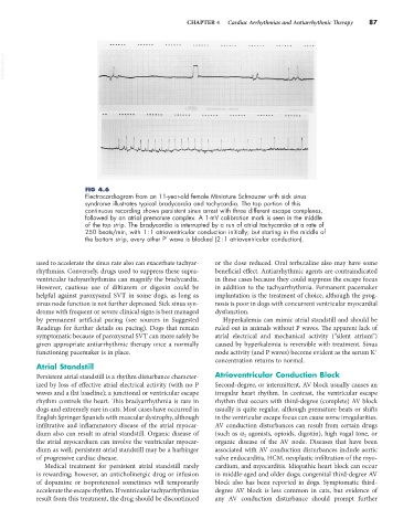
FIG 4.6
Electrocardiogram from an 11-year-old female Miniature Schnauzer with sick sinus
syndrome illustrates typical bradycardia and tachycardia. The top portion of this
continuous recording shows persistent sinus arrest with three different escape complexes,
followed by an atrial premature complex. A 1-mV calibration mark is seen in the middle
of the top strip. The bradycardia is interrupted by a run of atrial tachycardia at a rate of
250 beats/min, with 1 : 1 atrioventricular conduction initially; but starting in the middle of
the bottom strip, every other P′ wave is blocked (2 : 1 atrioventricular conduction).
used to accelerate the sinus rate also can exacerbate tachyar- or the dose reduced. Oral terbutaline also may have some
rhythmias. Conversely, drugs used to suppress these supra- beneficial effect. Antiarrhythmic agents are contraindicated
ventricular tachyarrhythmias can magnify the bradycardia. in these cases because they could suppress the escape focus
However, cautious use of diltiazem or digoxin could be in addition to the tachyarrhythmia. Permanent pacemaker
helpful against paroxysmal SVT in some dogs, as long as implantation is the treatment of choice, although the prog-
sinus node function is not further depressed. Sick sinus syn- nosis is poor in dogs with concurrent ventricular myocardial
drome with frequent or severe clinical signs is best managed dysfunction.
by permanent artificial pacing (see sources in Suggested Hyperkalemia can mimic atrial standstill and should be
Readings for further details on pacing). Dogs that remain ruled out in animals without P waves. The apparent lack of
symptomatic because of paroxysmal SVT can more safely be atrial electrical and mechanical activity (“silent atrium”)
given appropriate antiarrhythmic therapy once a normally caused by hyperkalemia is reversible with treatment. Sinus
+
functioning pacemaker is in place. node activity (and P waves) become evident as the serum K
concentration returns to normal.
Atrial Standstill
Persistent atrial standstill is a rhythm disturbance character- Atrioventricular Conduction Block
ized by loss of effective atrial electrical activity (with no P Second-degree, or intermittent, AV block usually causes an
waves and a flat baseline); a junctional or ventricular escape irregular heart rhythm. In contrast, the ventricular escape
rhythm controls the heart. This bradyarrhythmia is rare in rhythm that occurs with third-degree (complete) AV block
dogs and extremely rare in cats. Most cases have occurred in usually is quite regular, although premature beats or shifts
English Springer Spaniels with muscular dystrophy, although in the ventricular escape focus can cause some irregularities.
infiltrative and inflammatory disease of the atrial myocar- AV conduction disturbances can result from certain drugs
dium also can result in atrial standstill. Organic disease of (such as α 2 agonists, opioids, digoxin), high vagal tone, or
the atrial myocardium can involve the ventricular myocar- organic disease of the AV node. Diseases that have been
dium as well; persistent atrial standstill may be a harbinger associated with AV conduction disturbances include aortic
of progressive cardiac disease. valve endocarditis, HCM, neoplastic infiltration of the myo-
Medical treatment for persistent atrial standstill rarely cardium, and myocarditis. Idiopathic heart block can occur
is rewarding; however, an anticholinergic drug or infusion in middle-aged and older dogs; congenital third-degree AV
of dopamine or isoproterenol sometimes will temporarily block also has been reported in dogs. Symptomatic third-
accelerate the escape rhythm. If ventricular tachyarrhythmias degree AV block is less common in cats, but evidence of
result from this treatment, the drug should be discontinued any AV conduction disturbance should prompt further