
Page 116 - Small Animal Internal Medicine, 6th Edition
P. 116
88 PART I Cardiovascular System Disorders
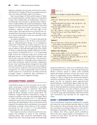
diagnostic evaluation, because most cases have been associ- BOX 4.4
ated with HCM. Complete AV block occasionally is found in
VetBooks.ir older cats without detectable organic heart disease. Formulas to Calculate Constant-Rate Infusion
Type I second-degree AV block and first-degree AV block
Method 1
are associated frequently with high vagal tone or drug effects
in dogs. These animals often are asymptomatic; exercise or (Allows for “fine-tuning” fluid and drug administration
anticholinergic drug (atropine or glycopyrrolate) adminis- rate)
tration usually abolishes the conduction disturbance. High- Determine desired drug infusion rate: µg/kg/min × kg
grade second-degree AV block (many blocked P waves) and body weight = µg/min (A)
complete (3°) heart block usually cause lethargy, exercise Determine desired fluid infusion rate: mL/hour ÷ 60 =
mL/min (B)
intolerance, weakness, syncope, and other signs of low (A) ÷ (B) = µg/min ÷ mL/min = µg drug/mL of fluid
cardiac output. These signs become severe at heart rates con- Convert from µg to mg of drug needed (1 µg =
sistently below 40 beats/min in dogs. CHF develops second- 0.001 mg)
ary to chronic bradycardia in some cases, especially if other Mg drug/mL fluid × mL of fluid in bag (or bottle, etc.) =
cardiac disease is present. mg of drug to add to the fluid container
An atropine challenge test (p. 97) is used to determine the
degree of vagal influence on the AV block. Oral anticholin- Method 2
ergic (or sympathomimetic) therapy can be attempted in (For total dose over a 6-hour period, must also calculate
symptomatic animals that are atropine-responsive (see Fig. fluid volume and administration rate)
4.5). However, atropine and oral anticholinergic therapy Total dose in mg to infuse over a 6-hour period = body
often is ineffective, especially with high-grade second-degree weight (kg) × dose (µg/kg/min) × 0.36
AV block, so artificial pacing usually is indicated. An emer- Method 3 (for Lidocaine)
gency infusion of dopamine (see Box 3.1) or isoproterenol (Faster but less helpful if fluid rate is important or if fine
might increase the ventricular escape rate in animals with drug-dosage adjustments are necessary)
high-grade second- or third-degree block, although it may For CRI of 44 µg/kg/min of lidocaine, add 25 mL of 2%
also provoke ventricular tachyarrhythmias. Oral isoprotere- lidocaine to 250 mL of D 5 W
nol usually is ineffective. A thorough cardiac workup is indi- Infuse at 0.25 mL/25 lb of body weight/min
cated before permanent artificial pacemaker implantation
because some underlying diseases (progressive myocardial
disease, endocarditis) are associated with a poor prognosis,
even after pacing. Temporary transvenous pacing could be antagonists (β-blockers), which act by inhibiting the effects
attempted for 1 to 2 days in questionable cases to assess the of catecholamines on the heart. Class III drugs prolong the
animal’s response to a normal heart rate before permanent effective refractory period of cardiac action potentials
pacemaker surgery is performed. More information on without decreasing conduction velocity; they may be most
pacing therapy is found in Suggested Readings. effective in suppressing reentrant arrhythmias and in pre-
venting VF. Class IV drugs are the calcium entry blockers;
ventricular arrhythmias usually are not responsive to these
ANTIARRHYTHMIC AGENTS agents, but they are important against supraventricular
tachyarrhythmias. Antiarrhythmic agents within this classi-
Antiarrhythmic drugs can act by slowing the rate of a tachy- fication scheme are contraindicated in animals with com-
cardia, terminating a reentrant arrhythmia, or preventing plete AV block and should be used only cautiously in animals
abnormal impulse formation or conduction. These effects with sinus bradycardia, sick sinus syndrome, and first- or
occur through modulation of tissue electrophysiologic second-degree AV block. Sometimes a designation of Class
properties and/or autonomic nervous system effects. The V is used for drugs with antiarrhythmic effects that work by
traditional (Vaughan-Williams) antiarrhythmic drugs are other mechanisms than described for the original four
classified according to their main electrophysiologic effects classes.
on cardiac cell action potentials (Table 4.1). Although this
classification system has several shortcomings (for example, CLASS I ANTIARRHYTHMIC DRUGS
+
some drugs with antiarrhythmic effects are excluded, several Class I antiarrhythmic drugs block membrane Na channels
drugs have the multiclass effects, and attention to ion channel and depress the action potential upstroke (phase 0), which
mechanisms is lacking), clinical reference to this classifica- slows conduction velocity along the cardiac cells. They have
tion persists. See Table 4.2 and Box 4.4 for antiarrhythmic been subclassified according to differences in other electro-
drug dosages and CRI calculation methods. physiologic characteristics. These differences (see Table 4.1)
Class I agents tend to slow conduction and decrease auto- can influence their efficacy against specific arrhythmias.
+
maticity and excitability by means of their membrane- Most of the class I agents depend on extracellular K con-
stabilizing effects; older ventricular antiarrhythmic drugs centration for their effects, and they lose effectiveness in
belong to this class. Class II drugs include the β-adrenergic patients with hypokalemia.