
Page 1196 - Small Animal Internal Medicine, 6th Edition
P. 1196
1168 PART IX Nervous System and Neuromuscular Disorders
(e.g., tick paralysis, botulism, acute fulminating myasthenia
gravis) using clinical features and (when available) electro-
VetBooks.ir diagnostic testing (see Table 66.2). Owners should be ques-
tioned about any possible inciting event or exposure 7 to 14
days earlier. Systemic evaluation including routine blood-
work, urinalysis, thoracic radiographs, and abdominal ultra-
sound are usually recommended. Normal cranial nerve and
esophageal function and the presence of hyperesthesia and
rapidly progressive muscle atrophy make ACP most likely.
A When performed 6 or more days after the onset of paresis,
electromyography reliably reveals diffuse denervation (fibril-
lation potentials and positive sharp waves), a finding not
expected with NMJ disorders. Additional EMG features
described in the Suggested Readings indicate a motor axo-
nopathy and demyelination that is most pronounced in the
ventral nerve roots and proximal nerves. Lumbar CSF analy-
sis typically reveals a normal cell count and distribution as
well as an increase in CSF protein, particularly 7 days or
more into clinical signs. In cases where there is no history of
potential raccoon exposure or recent vaccination, diagnostic
testing for an inciting cause (Toxoplasma, Neospora) should
be considered. Definitive diagnosis can also be established
B by nerve biopsy, but this is rarely necessary.
Treatment
No specific treatment exists for ACP. During the initial pro-
gressive phase, dogs must be monitored for respiratory com-
promise, especially if they have had a rapid progression to
recumbency. Signs typically stabilize after 5 to 10 days, after
which patients can usually be managed with supportive care
at home. They may require assistance in sitting up to eat and
drink. If possible, they should be kept on an air mattress,
waterbed, lounge chair, or bed of straw and turned periodi-
cally to prevent lung atelectasis and pressure sores. Gluco-
corticoid treatment is not beneficial, and may actually slow
recovery, but treatment with human intravenous immuno-
C globulin (IVIG, 0.5 g/kg slow intravenous [IV] infusion
q24h for 4 doses) (Sandoglobulin [Behring]) may help speed
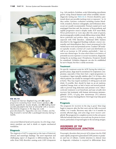
FIG 66.14 recovery.
A 4-year-old German Shepherd dog with (A) rapidly
progressive ascending lower motor neuron paralysis, (B) Prognosis
severe appendicular muscle atrophy, and (C) healing facial
wounds presumed to be from an encounter with a raccoon. The prognosis for recovery in the dog is good. Most dogs
The tentative diagnosis in this dog was acute begin to improve after the first week and are fully recovered
polyradiculoneuritis. Supportive care was initiated, and the within 3 to 4 weeks. Recovery may take 4 to 6 months in
dog returned to normal after a prolonged recovery lasting 3 severely affected dogs, and some dogs never recover com-
months. pletely. The prognosis for complete recovery in the cat is poor.
Affected animals that have recovered may be prone to recur-
rences, particularly if exposed again to the initiating antigen.
concurrent bilateral facial nerve paresis. In a few dogs, respi-
ratory paralysis can lead to death or require mechanical
ventilation. DISORDERS OF THE
NEUROMUSCULAR JUNCTION
Diagnosis
The diagnosis of ACP is suspected on the basis of historical, Presynaptic disorders that prevent ACh release into the NMJ
clinical, and neurologic findings. The most important and cause rapidly progressive generalized LMN paresis or paraly-
challenging aspect of diagnosis is differentiating this dis- sis and loss of reflexes. Careful attention to clinical clues
order from NMJ disorders causing acute LMN tetraparesis or diagnostic evaluation is required to differentiate these