Page 162 - Small Animal Internal Medicine, 6th Edition
P. 162
134    PART I   Cardiovascular System Disorders


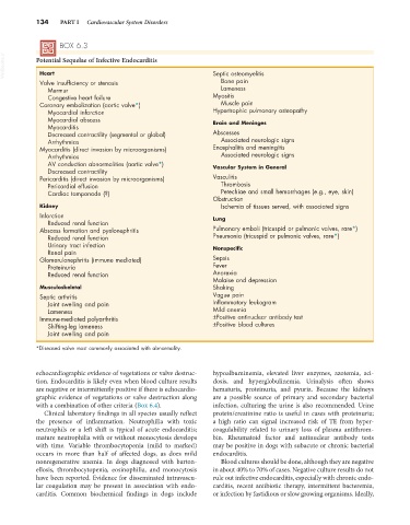
                   BOX 6.3
  VetBooks.ir  Potential Sequelae of Infective Endocarditis      Septic osteomyelitis

             Heart
             Valve insufficiency or stenosis                       Bone pain
               Murmur                                              Lameness
               Congestive heart failure                          Myositis
             Coronary embolization (aortic valve*)                 Muscle pain
               Myocardial infarction                             Hypertrophic pulmonary osteopathy
               Myocardial abscess                                Brain and Meninges
               Myocarditis
               Decreased contractility (segmental or global)     Abscesses
               Arrhythmias                                         Associated neurologic signs
             Myocarditis (direct invasion by microorganisms)     Encephalitis and meningitis
               Arrhythmias                                         Associated neurologic signs
               AV conduction abnormalities (aortic valve*)       Vascular System in General
               Decreased contractility
             Pericarditis (direct invasion by microorganisms)    Vasculitis
               Pericardial effusion                                Thrombosis
               Cardiac tamponade (?)                               Petechiae and small hemorrhages (e.g., eye, skin)
                                                                 Obstruction
             Kidney                                                Ischemia of tissues served, with associated signs
             Infarction                                          Lung
               Reduced renal function
             Abscess formation and pyelonephritis                Pulmonary emboli (tricuspid or pulmonic valves, rare*)
               Reduced renal function                            Pneumonia (tricuspid or pulmonic valves, rare*)
               Urinary tract infection                           Nonspecific
               Renal pain
             Glomerulonephritis (immune mediated)                Sepsis
               Proteinuria                                       Fever
               Reduced renal function                            Anorexia
                                                                 Malaise and depression
             Musculoskeletal                                     Shaking
             Septic arthritis                                    Vague pain
               Joint swelling and pain                           Inflammatory leukogram
               Lameness                                          Mild anemia
             Immune-mediated polyarthritis                       ±Positive antinuclear antibody test
               Shifting-leg lameness                             ±Positive blood cultures
               Joint swelling and pain
            *Diseased valve most commonly associated with abnormality.


            echocardiographic evidence of vegetations or valve destruc-  hypoalbuminemia, elevated liver enzymes, azotemia, aci-
            tion. Endocarditis is likely even when blood culture results   dosis, and hyperglobulinemia. Urinalysis often shows
            are negative or intermittently positive if there is echocardio-  hematuria, proteinuria, and pyuria. Because the kidneys
            graphic evidence of vegetations or valve destruction along   are a possible source of primary and secondary bacterial
            with a combination of other criteria (Box 6.4).      infection, culturing the urine is also recommended. Urine
              Clinical laboratory findings in all species usually reflect   protein/creatinine ratio is useful in cases with proteinuria;
            the presence of inflammation. Neutrophilia with toxic   a  high  ratio  can  signal  increased risk  of TE  from  hyper-
            neutrophils or a left shift is typical of acute endocarditis;   coagulability  related  to  urinary  loss  of  plasma  antithrom-
            mature neutrophilia with or without monocytosis develops   bin. Rheumatoid factor and antinuclear antibody tests
            with time. Variable thrombocytopenia (mild to marked)   may be positive in dogs with subacute or chronic bacterial
            occurs in more than half of affected dogs, as does mild   endocarditis.
            nonregenerative anemia. In dogs diagnosed with barton-  Blood cultures should be done, although they are negative
            ellosis, thrombocytopenia, eosinophilia, and monocytosis   in about 40% to 70% of cases. Negative culture results do not
            have been reported. Evidence for disseminated intravascu-  rule out infective endocarditis, especially with chronic endo-
            lar coagulation may be present in association with endo-  carditis, recent antibiotic therapy, intermittent bacteremia,
            carditis. Common biochemical findings in dogs include   or infection by fastidious or slow growing organisms. Ideally,

