
Page 164 - Small Animal Internal Medicine, 6th Edition
P. 164
136 PART I Cardiovascular System Disorders
VetBooks.ir
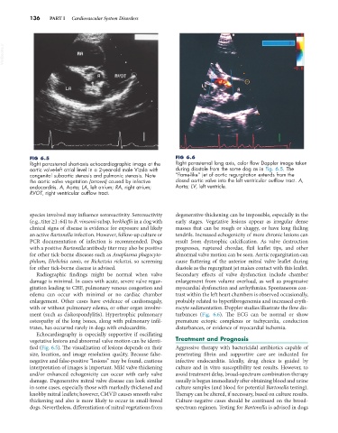
FIG 6.5 FIG 6.6
Right parasternal short-axis echocardiographic image at the Right parasternal long axis, color flow Doppler image taken
aortic valve-left atrial level in a 2-year-old male Vizsla with during diastole from the same dog as in Fig. 6.5. The
congenital subaortic stenosis and pulmonic stenosis. Note “flame-like” jet of aortic regurgitation extends from the
the aortic valve vegetation (arrows) caused by infective closed aortic valve into the left ventricular outflow tract. A,
endocarditis. A, Aorta; LA, left atrium; RA, right atrium; Aorta; LV, left ventricle.
RVOT, right ventricular outflow tract.
species involved may influence seroreactivity. Seroreactivity degenerative thickening can be impossible, especially in the
(e.g., titer ≥1 : 64) to B. vinsonii subsp. berkhoffii in a dog with early stages. Vegetative lesions appear as irregular dense
clinical signs of disease is evidence for exposure and likely masses that can be rough or shaggy, or have long flailing
an active Bartonella infection. However, follow-up culture or tendrils. Increased echogenicity of more chronic lesions can
PCR documentation of infection is recommended. Dogs result from dystrophic calcification. As valve destruction
with a positive Bartonella antibody titer may also be positive progresses, ruptured chordae, flail leaflet tips, and other
for other tick-borne diseases such as Anaplasma phagocyto- abnormal valve motion can be seen. Aortic regurgitation can
philum, Ehrlichia canis, or Rickettsia rickettsi, so screening cause fluttering of the anterior mitral valve leaflet during
for other tick-borne disease is advised. diastole as the regurgitant jet makes contact with this leaflet.
Radiographic findings might be normal when valve Secondary effects of valve dysfunction include chamber
damage is minimal. In cases with acute, severe valve regur- enlargement from volume overload, as well as progressive
gitation leading to CHF, pulmonary venous congestion and myocardial dysfunction and arrhythmias. Spontaneous con-
edema can occur with minimal or no cardiac chamber trast within the left heart chambers is observed occasionally,
enlargement. Other cases have evidence of cardiomegaly, probably related to hyperfibrogenemia and increased eryth-
with or without pulmonary edema, or other organ involve- rocyte sedimentation. Doppler studies illustrate the flow dis-
ment (such as diskospondylitis). Hypertrophic pulmonary turbances (Fig. 6.6). The ECG can be normal or show
osteopathy of the long bones, along with pulmonary infil- premature ectopic complexes or tachycardia, conduction
trates, has occurred rarely in dogs with endocarditis. disturbances, or evidence of myocardial ischemia.
Echocardiography is especially supportive if oscillating
vegetative lesions and abnormal valve motion can be identi- Treatment and Prognosis
fied (Fig. 6.5). The visualization of lesions depends on their Aggressive therapy with bactericidal antibiotics capable of
size, location, and image resolution quality. Because false- penetrating fibrin and supportive care are indicated for
negative and false-positive “lesions” may be found, cautious infective endocarditis. Ideally, drug choice is guided by
interpretation of images is important. Mild valve thickening culture and in vitro susceptibility test results. However, to
and/or enhanced echogenicity can occur with early valve avoid treatment delay, broad-spectrum combination therapy
damage. Degenerative mitral valve disease can look similar usually is begun immediately after obtaining blood and urine
in some cases, especially those with markedly thickened and culture samples (and blood for potential Bartonella testing).
knobby mitral leaflets; however, CMVD causes smooth valve Therapy can be altered, if necessary, based on culture results.
thickening and also is more likely to occur in small-breed Culture-negative cases should be continued on the broad-
dogs. Nevertheless, differentiation of mitral vegetations from spectrum regimen. Testing for Bartonella is advised in dogs