
Page 171 - Small Animal Internal Medicine, 6th Edition
P. 171
CHAPTER 7 Myocardial Diseases of the Dog 143
VetBooks.ir
A B
C D
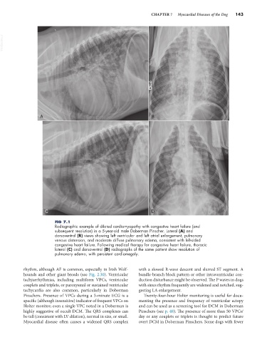
FIG 7.1
Radiographic example of dilated cardiomyopathy with congestive heart failure (and
subsequent resolution) in a 5-year-old male Doberman Pinscher. Lateral (A) and
dorsoventral (B) views showing left ventricular and left atrial enlargement, pulmonary
venous distension, and moderate diffuse pulmonary edema, consistent with left-sided
congestive heart failure. Following medical therapy for congestive heart failure, thoracic
lateral (C) and dorsoventral (D) radiographs of the same patient show resolution of
pulmonary edema, with persistent cardiomegaly.
rhythm, although AF is common, especially in Irish Wolf- with a slowed R-wave descent and slurred ST segment. A
hounds and other giant breeds (see Fig. 2.30). Ventricular bundle-branch block pattern or other intraventricular con-
tachyarrhythmias, including multiform VPCs, ventricular duction disturbance might be observed. The P waves in dogs
couplets and triplets, or paroxysmal or sustained ventricular with sinus rhythm frequently are widened and notched, sug-
tachycardia are also common, particularly in Doberman gesting LA enlargement.
Pinschers. Presence of VPCs during a 5-minute ECG is a Twenty-four-hour Holter monitoring is useful for docu-
specific (although insensitive) indicator of frequent VPCs on menting the presence and frequency of ventricular ectopy
Holter monitor; even a single VPC noted in a Doberman is and can be used as a screening tool for DCM in Doberman
highly suggestive of occult DCM. The QRS complexes can Pinschers (see p. 48). The presence of more than 50 VPCs/
be tall (consistent with LV dilation), normal in size, or small. day or any couplets or triplets is thought to predict future
Myocardial disease often causes a widened QRS complex overt DCM in Doberman Pinschers. Some dogs with fewer