
Page 172 - Small Animal Internal Medicine, 6th Edition
P. 172
144 PART I Cardiovascular System Disorders
than 50 VPCs/day on initial evaluation also develop DCM Echocardiography is used to screen for occult myocardial
after several years. The frequency and complexity of ven- disease also. Screening is complicated by the fact that appar-
VetBooks.ir tricular tachyarrhythmias appear to be negatively correlated ently healthy Doberman Pinschers, Greyhounds, and some
other athletic dogs can have slightly reduced fractional
with fractional shortening; sustained ventricular tachycardia
has been associated with increased risk of sudden death.
normal for most breeds. For asymptomatic Doberman Pin-
Variability in the number of VPCs between repeated Holter shortening compared with what is generally considered
recordings in the same dog can be high (up to 85%). If avail- schers, the following echocardiographic criteria suggest
able, the technique of signal-averaged electrocardiography occult DCM with a high risk for overt disease within 2 to 3
can reveal the presence of ventricular late potentials, which years: LVIDs greater than 4.6 cm (in dogs ≤42 kg) or greater
could suggest an increased risk for sudden death in Dober- than 5.0 cm (in dogs >42 kg), LVIDs greater than 3.8 cm,
man Pinschers with occult DCM. mitral valve E point–septal separation greater than 0.9 cm,
or VPCs during initial examination (LVID, left ventricular
ECHOCARDIOGRAPHY internal diameter; d, diastole; s, systole).
Echocardiography is used to definitively diagnose DCM
(and differentiate from pericardial effusion or chronic mitral Clinicopathologic Findings
valve disease), assess severity of systolic dysfunction, and Circulating concentrations of the natriuretic peptides (B-type
document degree of cardiac chamber enlargement. Dilated natriuretic peptide [BNP] and atrial natriuretic peptide
cardiac chambers and poor ventricular systolic wall motion [ANP]) and cardiac troponin are elevated in Doberman
are characteristic findings in dogs with DCM (Fig. 7.2). In Pinschers with occult DCM, and levels of these biomarkers
severe cases only, minimal wall motion is evident. Left heart rise as disease progresses and CHF develops. Among these
enlargement predominates, although all chambers are usually biomarkers, NT-proBNP appears to have the best sensitivity
affected to some degree. Echocardiographic indices of LV and specificity for detecting occult DCM, particularly when
systolic function are decreased, including fractional shorten- echocardiographic abnormalities are present. However, NT-
ing, fractional area change, and ejection fraction. LV systolic proBNP has wide biologic variability in normal dogs and is
(as well as diastolic) dimension is increased compared with relatively insensitive for detecting occult DCM when ventric-
normal ranges for the breed; the LV appears more spherical, ular arrhythmias precede echocardiographic changes. Thus
and mitral valve E point–septal separation is increased. LV the gold standard screening regimen for detecting occult
free-wall and septal thicknesses are normal to decreased. The DCM in individual dogs is combined Holter monitoring
calculated end-systolic volume index (see p. 25) typically is and echocardiography. In high-volume screening situations,
2
greater than 80 mL/m in dogs with overt DCM (<30 mL/ a combination of Holter monitoring and NT-proBNP testing
m is considered normal). Evidence for abnormal diastolic could be considered. Genetic screening is recommended in
2
function also can be found in dogs with advanced disease. Doberman Pinschers intended for breeding.
Mild to moderate centrally directed AV valve regurgitation
usually is seen with Doppler echocardiography (Fig. 7.3).
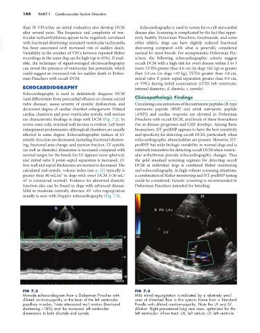
FIG 7.2 FIG 7.3
M-mode echocardiogram from a Doberman Pinscher with Mild mitral regurgitation is indicated by a relatively small
dilated cardiomyopathy at the level of the left ventricular area of disturbed flow in this systolic frame from a Standard
papillary muscles. Note attenuated wall motion (fractional Poodle with dilated cardiomyopathy. Note the LA and LV
shortening ~18%) and the increased left ventricular dilation. Right parasternal long axis view, optimized for the
dimensions in both diastole and systole. left ventricular inflow tract. LA, Left atrium; LV, left ventricle.