
Page 197 - Small Animal Internal Medicine, 6th Edition
P. 197
CHAPTER 8 Myocardial Diseases of the Cat 169
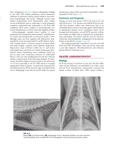
heart enlargement (Fig. 8.7). Typical radiographic findings classification scheme often are termed “unclassified” cardio-
in cats with CHF include pulmonary venous distension, myopathy (UCM) (see p. 171).
VetBooks.ir infiltrates of pulmonary edema, pleural effusion, and some- Treatment and Prognosis
times hepatomegaly and ascites. Although normal sinus
rhythm predominates, ECG abnormalities often include
with HCM (see p. 164). Because cats with RCM do not typi-
various arrhythmias such as ventricular or atrial premature Therapy for acute and chronic CHF is the same as for cats
complexes, supraventricular tachycardia, or AF. Wide QRS cally have dynamic outflow tract obstruction, there is no
complexes, tall R waves, evidence of intraventricular con- contraindication to positive inotropic agents. Pimobendan is
duction disturbances, or wide P waves also might be evident. an appropriate therapy; severe cardiogenic shock can be
Echocardiography typically shows marked LA (and managed with dobutamine. As with HCM, atenolol or diltia-
sometimes RA) enlargement with normal LV wall thickness. zem usually are added only as treatment for tachyarrhyth-
LV systolic function generally is normal (FS usually > 25%), mias, particularly AF. Sotalol could be used for refractory
although some cats have regional wall dysfunction. End- ventricular tachyarrhythmias. Management of thromboem-
stage RCM can be associated with LV and RV dilation. Pulse- bolism is described in Chapter 12, p. 227.
wave Doppler shows a restrictive pattern of mitral inflow, The prognosis generally is guarded to poor for cats with
and tissue Doppler confirms severe diastolic dysfunction. RCM and CHF. Nevertheless, some cats survive more than
Hyperechoic areas of fibrosis within the LV wall and/or a year after diagnosis. Thromboembolism and refractory
endocardial areas sometimes are evident. Extraneous intra- pleural effusion commonly occur.
luminal echoes representing excess moderator bands are
seen in some cases. Sometimes, extensive LV endocardial
fibrosis, with scar tissue bridging between the free-wall and DILATED CARDIOMYOPATHY
septum, constricts part of the ventricular chamber. An intra-
cardiac thrombus might be found, usually in the left auricle Etiology
or LA but occasionally in the LV. Mild mitral or tricuspid DCM has become uncommon in cats since the late 1980s,
regurgitation often is present. As previously discussed, dif- when taurine deficiency was identified as its major cause,
ferentiation between RCM and end-stage remodeled HCM and pet food manufacturers subsequently increased the
can be challenging. Cases that do not fit within the typical taurine content of feline diets. Other factors besides a
A B
FIG 8.7
Lateral (A) and dorsoventral (B) radiographs from a domestic shorthair cat with restrictive
cardiomyopathy show marked left atrial enlargement and prominent pulmonary vessels.