
Page 192 - Small Animal Internal Medicine, 6th Edition
P. 192
164 PART I Cardiovascular System Disorders
inhibitor (ACEI), or spironolactone have been performed,
but clear benefit from any of these interventions has yet to be
VetBooks.ir proven. With this in mind, some clinicians still suggest using
a β-blocker in cats with evidence of substantial dynamic LV
outflow obstruction or arrhythmias. In cats with marked,
nonobstructive myocardial hypertrophy, particularly with
evidence of myocardial fibrosis and remodeling, an ACEI
might be suggested. For cats with LA enlargement, especially
with spontaneous echocontrast, instituting antithrombotic
prophylaxis with clopidogrel is prudent (see Chapter 12).
Avoidance of stressful situations likely to cause persis-
tent tachycardia and reevaluation on a semiannual or annual
basis are usually advised. Secondary causes of myocardial
hypertrophy, such as systemic arterial hypertension and
hyperthyroidism, should be ruled out (or treated, if found).
CONGESTIVE HEART FAILURE
(STAGE C DISEASE)
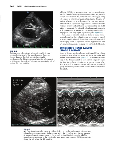
FIG 8.4
Right parasternal short-axis echocardiographic image Goals of therapy are to enhance ventricular filling, relieve
obtained at the level of the heart base in a 5-year-old congestion, control arrhythmias, minimize ischemia, and
female domestic shorthair cat with hypertrophic prevent thromboembolism (Box 8.1). Furosemide is used
cardiomyopathy. Note the massive left atrial enlargement only at the dosage needed to help control congestive signs
and thrombus (arrows) within the auricle. Ao, Aorta; LA, left
atrium; RV, right ventricle. for long-term therapy. Moderate to severe pleural effu-
sion is treated by thoracocentesis, with the cat restrained
gently in sternal position (and sedated with butorphanol
if needed).
A B
FIG 8.5
(A) Two-dimensional echo image in midsystole from a middle-aged domestic shorthair cat.
Echoes from the anterior mitral leaflet appear within the LV outflow tract (arrow) because
of abnormal systolic anterior (toward the septum) motion (SAM) of the valve. (B) The
M-mode echocardiogram at the mitral valve level also shows the mitral SAM (arrows). Ao,
Aorta; LA, left atrium; LV, left ventricle.