
Page 190 - Small Animal Internal Medicine, 6th Edition
P. 190
162 PART I Cardiovascular System Disorders
occur more commonly in cats with HCM compared with
cats without structural heart disease. Although LV changes
VetBooks.ir predominate, 30% to 50% of cats with HCM can have at
least segmental RV hypertrophy, and some cats have RA
dilation as well.
LA enlargement in cats with HCM can range from mild
to marked (see Chapter 2 and Figs. 8.3, A and D, and 8.4).
Prominent LA enlargement is expected in cats with clinical
signs of CHF. Spontaneous echocontrast (swirling, smoky
echoes) is visible within the enlarged LA of some cats. This
is thought to result from blood stasis with cellular aggrega-
tions, and to be a harbinger of thromboembolism. A throm-
bus occasionally is visualized within the LA, usually in the
auricle (see Fig. 8.4).
Cats with dynamic LV outflow tract obstruction often
have SAM of the mitral valve (Fig. 8.5) or premature closure
of the aortic valve leaflets on M-mode scans. Abnormalities
of the mitral valve apparatus, including increased papillary
muscle hypertrophy and anterior mitral leaflet length, have
been associated with SAM and severity of dynamic LV
outflow obstruction. Doppler modalities can demonstrate
mitral regurgitation and LV outflow turbulence (Fig. 8.6).
Continuous-wave Doppler can be used to demonstrate high-
velocity and late-peaking blood flow through the LV outflow
tract, confirming the dynamic obstruction. The left apical
five-chamber view may be most useful.
Doppler-derived estimates of diastolic function are now
routinely employed to define disease characteristics in HCM.
Pulsed wave (PW) Doppler may show a delayed relaxation
mitral inflow pattern (E wave:A wave velocity < 1) or evi-
dence for more advanced diastolic dysfunction. Prolonged
isovolumic relaxation time is associated with early diastolic
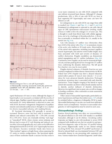
FIG 8.2 dysfunction. Tissue Doppler imaging of the lateral or septal
Electrocardiogram from a cat with hypertrophic
cardiomyopathy showing occasional ventricular premature mitral valve annulus can detect reduced early annular motion
complexes and a left axis deviation. Leads I, II, III, at in diastole, another hallmark of diastolic dysfunction.
25 mm/sec. 1 cm = 1 mV. However, the rapid heart rate in many cats, as well as changes
in loading conditions, often confounds accurate assessment
of diastolic function.
septal thicknesses of 8 mm or more, although the degree of Other causes of myocardial hypertrophy, particularly sys-
hypertrophy is not necessarily correlated with the severity of temic hypertension and hyperthyroidism (see p. 167), should
clinical signs. Papillary muscle hypertrophy can be marked, be excluded before a diagnosis of idiopathic HCM is made.
and systolic LV cavity obliteration is observed in some cats Myocardial thickening in cats also can result from infiltrative
with HCM. Increased echogenicity (brightness) of papillary disease (such as lymphoma). Variation in myocardial echo-
muscles and subendocardial areas is thought to be a marker genicity or wall irregularities may be noted in such cases.
for chronic myocardial ischemia, with resulting fibrosis.
LV fractional shortening (FS) generally is normal to Clinicopathologic Findings
increased. However, some cats have mild to moderate LV Routine clinical pathology tests often are noncontributory.
dilation and reduced contractility (FS ≈ 23%-29%; normal The pleural effusion in cats with CHF usually is a modified
FS is 35%-65%). Some cats eventually develop “end-stage” transudate, although it can be chylous. Circulating cardiac
or “remodeled” HCM, where chronic severe ischemia and troponins are higher in cats with moderate to severe HCM
fibrosis lead to areas of LV wall thinning and reduced con- compared with unaffected cats, but low sensitivity and speci-
tractility. Echocardiographically, such cases can be difficult to ficity limit diagnostic value of this test. NT-proBNP testing
distinguish from RCM or DCM, as LV hypertrophy becomes has proven diagnostically useful in two clinical settings
less dramatic. Excessive moderator bands (also known as involving HCM. First, elevated NT-proBNP (performed on
“false tendons”) appear as bright linear echoes spanning the blood or pleural fluid) can discriminated between CHF and
LV cavity in various configurations. The functional signifi- noncardiac disease in cats presenting with respiratory dis-
cance of these moderator bands is unclear, but they seem to tress. Various studies have identified diagnostic cutoff values