
Page 191 - Small Animal Internal Medicine, 6th Edition
P. 191
CHAPTER 8 Myocardial Diseases of the Cat 163
VetBooks.ir
A B
C D
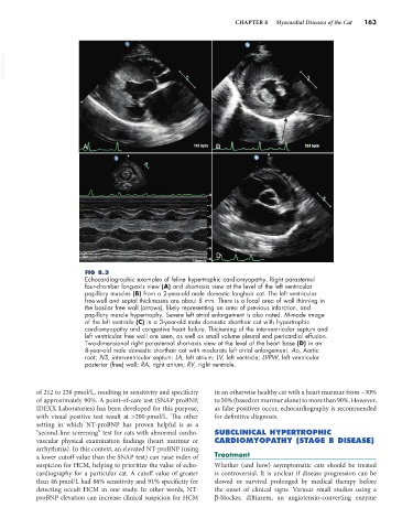
FIG 8.3
Echocardiographic examples of feline hypertrophic cardiomyopathy. Right parasternal
four-chamber long-axis view (A) and short-axis view at the level of the left ventricular
papillary muscles (B) from a 2-year-old male domestic longhair cat. The left ventricular
free-wall and septal thicknesses are about 8 mm. There is a focal area of wall thinning in
the basilar free wall (arrows), likely representing an area of previous infarction, and
papillary muscle hypertrophy. Severe left atrial enlargement is also noted. M-mode image
of the left ventricle (C) in a 3-year-old male domestic shorthair cat with hypertrophic
cardiomyopathy and congestive heart failure. Thickening of the interventricular septum and
left ventricular free wall are seen, as well as small volume pleural end pericardial effusion.
Two-dimensional right parasternal short-axis view at the level of the heart base (D) in an
8-year-old male domestic shorthair cat with moderate left atrial enlargement. Ao, Aortic
root; IVS, interventricular septum; LA, left atrium; LV, left ventricle; LVPW, left ventricular
posterior (free) wall; RA, right atrium; RV, right ventricle.
of 212 to 258 pmol/L, resulting in sensitivity and specificity in an otherwise healthy cat with a heart murmur from ~30%
of approximately 90%. A point-of-care test (SNAP proBNP, to 50% (based on murmur alone) to more than 90%. However,
IDEXX Laboratories) has been developed for this purpose, as false positives occur, echocardiography is recommended
with visual positive test result at >200 pmol/L. The other for definitive diagnosis.
setting in which NT-proBNP has proven helpful is as a
“second-line screening” test for cats with abnormal cardio- SUBCLINICAL HYPERTROPHIC
vascular physical examination findings (heart murmur or CARDIOMYOPATHY (STAGE B DISEASE)
arrhythmia). In this context, an elevated NT-proBNP (using
a lower cutoff value than the SNAP test) can raise index of Treatment
suspicion for HCM, helping to prioritize the value of echo- Whether (and how) asymptomatic cats should be treated
cardiography for a particular cat. A cutoff value of greater is controversial. It is unclear if disease progression can be
than 46 pmol/L had 86% sensitivity and 91% specificity for slowed or survival prolonged by medical therapy before
detecting occult HCM in one study. In other words, NT- the onset of clinical signs. Various small studies using a
proBNP elevation can increase clinical suspicion for HCM β-blocker, diltiazem, an angiotensin-converting enzyme