
Page 225 - Small Animal Internal Medicine, 6th Edition
P. 225
CHAPTER 10 Pulmonary Hypertension and Heartworm Disease 197
ELECTROCARDIOGRAPHY from platelet consumption in the pulmonary arterial system,
ECG findings are usually normal, although advanced disease especially after adulticide treatment. DIC also develops in
VetBooks.ir can cause a right axis deviation or arrhythmias, as with other some dogs with advanced disease. The immune response to
HWs produces a polyclonal gammopathy. Mild to moderate
causes of severe PAH (see p. 192).
ECHOCARDIOGRAPHY elevations in liver enzyme activity may be seen, especially in
cases of right-sided CHF (passive congestion of the liver).
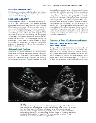
Echocardiographic findings in dogs with advanced HWD Azotemia, either prerenal or secondary to immune-complex
are as for other causes of severe PAH, with the additional glomerulonephritis, occasionally occurs. Proteinuria is
possibility of visualizing HWs in the right heart. Although found in 10% to 30% of affected dogs and is more likely with
HWs located in peripheral pulmonary arteries cannot be advanced disease; hypoalbuminemia can develop in severely
seen echocardiographically, worms within the main pulmo- affected dogs. If hypoalbuminemia or proteinuria is detected,
nary artery and its bifurcation, RV, RA, or vena cavae appear a urine protein-creatinine ratio (UPC) is advised. Tracheal
as small, bright parallel echoes (Fig. 10.3). Suspected caval washings from coughing HW-infected dogs usually indicate
syndrome can be quickly confirmed by echocardiography. eosinophilic inflammation.
Ascites or pleural or pericardial effusion accompanies sec-
ondary right-sided CHF. Color-flow Doppler imaging can
often reveal TR even when an audible murmur is absent. Treatment of Dogs With Heartworm Disease
Spectral Doppler measurement of maximum tricuspid (or
pulmonary) regurgitant jet velocity allows estimation of PREADULTICIDE EVALUATION
PAH severity (see p. 30). AND TREATMENT
As a general rule, adulticide treatment is recommended for
Clinicopathologic Findings dogs infected with HWs. Dogs who test positive for HW
Eosinophilia, basophilia, neutrophilia, and monocytosis are Ag should have a thorough history and physical examina-
common but inconsistent hematologic findings. Mild regen- tion. Pretreatment thoracic radiographs provide the best
erative anemia of inflammatory disease might be noted. overall assessment of pulmonary arterial and parenchymal
More severe anemia secondary to intravascular hemolysis disease status. The risk of postadulticide PTE is increased
occurs in caval syndrome. Thrombocytopenia can result in dogs with preexisting clinical and radiographic signs
A B
FIG 10.3
Echocardiographic images from a 4-year-old female spayed Beagle with caval syndrome.
Views from the right parasternal (A) four-chamber long-axis and (B) short-axis at the level
of the heart base are shown. There is enlargement of the right ventricle and main
pulmonary artery, with right ventricular hypertrophy. Many small, bright parallel echoes
are apparent in the body of the right ventricle (arrows), caused by a clump of heartworms
entangled in the tricuspid valve apparatus. Ao, Aortic root; LA, left atrium; LV, left
ventricle; PA, main pulmonary artery; RV, right ventricle.