
Page 220 - Small Animal Internal Medicine, 6th Edition
P. 220
192 PART I Cardiovascular System Disorders
ELECTROCARDIOGRAPHY underlying causes of PAH including pulmonary thrombo-
Electrocardiographic (ECG) findings often are normal, emboli and pulmonary fibrosis.
VetBooks.ir although severe PAH can cause a right axis deviation from Clinicopathologic Findings
RV enlargement. Tall P waves suggestive of right atrial
enlargement might also be found. Arrhythmias, such as ven-
seen in dogs with severe PAH. Arterial blood gas evaluation
tricular premature complexes (originating from the RV) or Increased red blood cell distribution width (RDW) often is
atrial fibrillation, can occur with advanced cor pulmonale. may show hypoxemia and hypercapnia. Other routine labo-
ratory test results (complete blood count [CBC], chemistry
ECHOCARDIOGRAPHY panel, urinalysis) will vary depending on the underlying
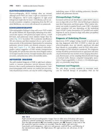
Echocardiographic findings in dogs with severe PAH include cause of PAH. Cardiac biomarkers (NT-proBNP and cardiac
RV and RA dilation, RV hypertrophy, flattening of the inter- troponin I) can be elevated in dogs with either precapillary
ventricular septum with paradoxical septal motion, a small or postcapillary PAH.
left heart, and pulmonary artery dilation (larger than the
aorta) (Fig. 10.2). Secondary TR or pulmonic insufficiency Diagnosis of Underlying Disease
(PI) are common, and their maximal velocity can be used to Heartworm (HW) antigen testing should be performed in
assess severity of the pulmonary hypertension by estimating any dog diagnosed with PAH. If HWD is ruled out and
pulmonary arterial systolic and diastolic pressures, respec- echocardiography does not identify significant left-sided
tively (see Chapter 2, p. 30). More advanced echocardio- heart disease as a postcapillary cause for PAH, other poten-
graphic indices suggestive of PAH include transpulmonic tial etiologies of PAH should be considered. This diagnostic
flow profile, RV systolic time intervals, tissue Doppler indices workup should be prioritized based on signalment and clini-
of tricuspid annulus motion, tricuspid annular plane systolic cal presentation but may include airway imaging (bronchos-
excursion, and pulmonary artery distensibility index. copy, fluoroscopy), airway sampling (bronchoalveolar lavage
or tracheal wash), thoracic computed tomography, assess-
ADVANCED IMAGING ment of hypercoagulability (D-dimers or thromboelastogra-
The gold standard diagnosis of PAH is right heart catheter- phy), or, potentially, lung biopsy.
ization to measure pulmonary arterial pressures directly;
however, this is rarely performed in clinical practice. Com- Treatment and Prognosis
puted tomography with contrast can confirm size and tortu- The only drugs currently available in veterinary medi-
osity of pulmonary arteries, and may be useful in diagnosing cine for directed therapy of precapillary PAH are the
A B
FIG 10.2
Right parasternal echocardiographic images from an older male Chihuahua with severe
pulmonary arterial hypertension secondary to chronic bronchopulmonary disease. Views
from the right parasternal (A) four-chamber long-axis and (B) short-axis at the level of the
left ventricular papillary muscles are provided. Note the severe right ventricular
hypertrophy and enlargement, right atrial enlargement, small left heart, and flattening of
the interventricular septum. RA, Right atrium; RV, right ventricle; LA, left atrium; LV, left
ventricle.