
Page 219 - Small Animal Internal Medicine, 6th Edition
P. 219
CHAPTER 10 Pulmonary Hypertension and Heartworm Disease 191
an example of postcapillary PAH; all other causes of PAH are with point of maximal intensity on the left hemithorax.
precapillary. Softer or right-sided heart murmurs raise index of suspicion
VetBooks.ir Clinical Findings for PAH. Dogs with left-sided CHF typically have sinus
tachycardia with heart rates of 150 to 160 beats per minute
because of sympathetic nervous system stimulation or may
Clinical signs of moderate to severe PAH include reduced
exercise tolerance, fatigue, persistent respiratory difficulty, have tachyarrhythmias such as ventricular premature com-
cough, and syncope. As these clinical signs overlap with plexes or atrial fibrillation. Dogs with PAH generally have a
common clinical signs of many primary respiratory diseases, sinus arrhythmia and/or relative sinus bradycardia related to
it often is challenging to determine whether clinical signs are elevated parasympathetic tone from underlying respiratory
directly attributable to PAH or to the underlying disease. disease.
Severe PAH also can lead to right heart remodeling (cor
pulmonale) and eventual right-sided congestive heart failure Diagnosis
(CHF), usually manifesting as ascites. Physical examination
findings could include cyanotic mucous membranes (at rest RADIOGRAPHY
or with exertion), a split S 2 heart sound, right-sided systolic Radiographic findings in patients with moderate to severe
heart murmur (of tricuspid regurgitation [TR]), and possibly PAH can include RV enlargement; main pulmonary artery
jugular venous distension and/or pulsation. Heart rate and dilation (“bulge” of the pulmonary trunk); and enlargement,
rhythm are usually normal; sinus arrhythmia and relative tortuosity, and blunting of lobar pulmonary arteries (Fig.
sinus bradycardia may reflect presence of underlying pulmo- 10.1) Caudal lobar arteries can be considered enlarged if
nary pathology causing elevated vagal tone. their width on dorsoventral (DV) or ventrodorsal (VD)
The clinical presentation of a dog with severe precapillary views is greater than the width of the proximal third rib.
PAH (respiratory distress, cough, syncope) closely mimics Occasionally, dogs with severe PAH have patchy alveolar
presentation of a dog with pulmonary edema secondary to infiltrates that resolve rapidly with sildenafil administration.
left-sided CHF. An additional confounding auscultatory These infiltrates are thought to represent a variant of non-
finding is pulmonary crackles, which are common in dogs cardiogenic pulmonary edema caused by regional nonuni-
with pulmonary edema but can also be present in dogs with formity in pulmonary capillary perfusion. Variable reactive
PAH secondary to pulmonary fibrosis or chronic pneumo- pulmonary arterial vasoconstriction causes some areas of
nia. Aspects of the physical examination can help differenti- lung to be overperfused compared with others, leading to
ate these two presentations before obtaining a more definitive focally high hydrostatic pressure and edema formation.
diagnosis with diagnostic imaging (thoracic radiographs and These alveolar infiltrates must be differentiated from cardio-
echocardiography). Dogs with left-sided CHF almost always genic pulmonary edema (caused by left-sided CHF), because
have loud systolic heart murmurs (grade IV/VI or louder) sildenafil is the preferred treatment.
A B
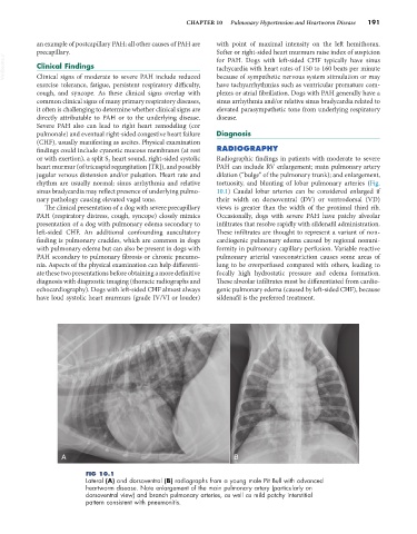
FIG 10.1
Lateral (A) and dorsoventral (B) radiographs from a young male Pit Bull with advanced
heartworm disease. Note enlargement of the main pulmonary artery (particularly on
dorsoventral view) and branch pulmonary arteries, as well as mild patchy interstitial
pattern consistent with pneumonitis.