
Page 215 - Small Animal Internal Medicine, 6th Edition
P. 215
CHAPTER 9 Pericardial Disease and Cardiac Tumors 187
relate to bleeding tumors (e.g., HSA) present in extracardiac masses are accentuated by the echolucent intracardiac blood
locations as well. surrounding them (Fig. 9.8). The left cranial parasternal
VetBooks.ir sounds (if large pericardial effusion is present) are common. transducer position is especially useful for evaluating the
Auscultatory findings vary. Arrhythmias or muffled heart
ascending aorta, right auricle, and surrounding structures.
Sometimes a murmur is caused by partial obstruction of
lesion can suggest the type of tumor, although cytologic or
intracardiac blood flow caused by the tumor mass, but The location and echocardiographic characteristics of a mass
murmurs associated with unrelated disease (for example, histopathologic evaluation is necessary for definitive diag-
chronic mitral valve disease) are more common. Conversely, nosis. HSA typically has variable echogenicity, with areas
auscultatory findings can be normal. that appear cystic (hypoechoic). Chemodectoma and other
heart base masses tend to have a more uniform soft tissue
Diagnosis echogenicity. Myocardial lymphoma also can have a mottled
Hematologic and serum biochemical tests generally are non- appearance with areas of varying echogenicity. Echocardio-
specific in dogs and cats with cardiac tumors; with flow graphic assessment of the tumor’s location, size, attachment
cytometry based hematology analyzers, neoplastic cells are (pedunculated or broad based), and extent (superficial or
frequently detected in the graphics of dogs with lymphoma deeply invading adjacent myocardium) may help in deter-
or malignant histiocytosis. Plasma cTnI concentrations are mining whether surgical resection or biopsy is possible.
likely to be elevated (>0.25 ng/mL) in dogs with cardiac HSA, Visualizing a suspected mass lesion in more than one echo-
compared with dogs with noncardiac HSA, other neoplasms, cardiographic plane helps verify it and prevent the misin-
or pericardial effusion not caused by HSA. Mild increases in terpretation of artifacts. Fine-needle aspirates for cytologic
serum alanine aminotransferase activity and azotemia might evaluation can be done under echocardiographic guidance
occur in dogs with CHF signs. HSA can be associated with in some cases. A discrete mass lesion often is not found with
a regenerative anemia, increased number of nucleated red mesothelioma.
blood cells and schistocytes (with or without acanthocytes), Pericardial fluid analysis is recommended, although
leukocytosis, and thrombocytopenia. Pleural and peritoneal definitive diagnosis of neoplasia usually cannot be made
fluids, if present, are usually modified transudates. based on cytologic findings alone (see p. 181). Cardiac lym-
Radiographic findings are quite variable. The cardiac sil- phoma or malignant histiocytosis is more likely to be diag-
houette may be normal or show an unusual bulge, a mass nosed on pericardial fluid cytology. Nevertheless, visualization
effect adjacent to the heart, or a globoid cardiac silhouette
compatible with pericardial effusion. Intrapericardial masses
usually are obscured by pericardial effusion. Caudal vena
caval distension, pleural effusion, and/or ascites commonly
occur with RV inflow or outflow obstruction. Dorsal devia-
tion of the trachea and increased perihilar opacity are seen
in some dogs with heart base tumors. Evidence of pulmo-
nary metastases is found with some primary or secondary
(metastatic) cardiac neoplasms; however, radiographic sen-
sitivity for detecting small pulmonary metastases is low. CT,
MRI, or other imaging techniques also can help in identify-
ing and defining the extent of cardiac tumors.
The ECG might suggest pericardial effusion (see p. 181).
Myocardial infiltration can provoke atrial or ventricular pre-
mature complexes or paroxysmal tachycardias. Likewise,
varying degrees of AV or intraventricular conduction block
and symptomatic bradycardia can develop from conduction
system infiltration. Intracardiac tumors that obstruct RV
outflow, causing RV systolic pressure overload and compen-
satory myocardial hypertrophy, can produce a right axis shift
and RV hypertrophy pattern on the ECG. Other chamber
enlargement or abnormal conduction patterns could result,
depending on tumor location and hemodynamic sequelae.
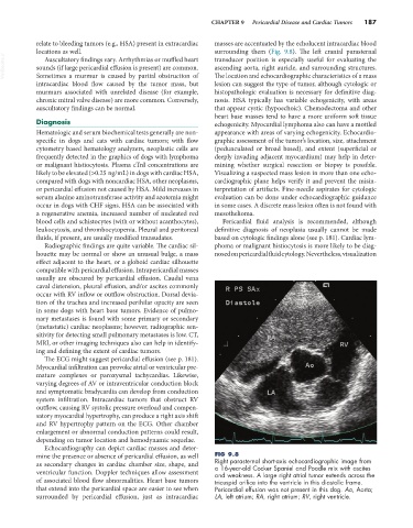
Echocardiography can depict cardiac masses and deter-
mine the presence or absence of pericardial effusion, as well FIG 9.8
as secondary changes in cardiac chamber size, shape, and Right parasternal short-axis echocardiographic image from
a 16-year-old Cocker Spaniel and Poodle mix with ascites
ventricular function. Doppler techniques allow assessment and weakness. A large right atrial tumor extends across the
of associated blood flow abnormalities. Heart base tumors tricuspid orifice into the ventricle in this diastolic frame.
that extend into the pericardial space are easier to see when Pericardial effusion was not present in this dog. Ao, Aorta;
surrounded by pericardial effusion, just as intracardiac LA, left atrium; RA, right atrium; RV, right ventricle.