
Page 464 - Small Animal Internal Medicine, 6th Edition
P. 464
436 PART III Digestive System Disorders
necessitates the use of commercial liquid diets (Table 28.2) or long hemostat or other device. Esophagostomy tubes do
instead of homemade gruels. The clinician should flush not cause gagging, a problem common with pharyngostomy
VetBooks.ir the tube with water after each feeding to prevent occlu- tubes.
Gastrostomy tubes bypass the mouth and esophagus in
sion. Long-term acceptance is typical, but rhinitis occurs in
some animals.
also be used when nasoesophageal, esophagostomy, or inter-
Some dogs and cats do not tolerate nasoesophageal tubes animals with a functional stomach and intestines. They can
and repeatedly pull them out. However, they are usually mittent gastric tubing is unacceptable. Vomiting is not a
effective for short-term therapy (e.g., 1-10 days), and some contraindication. This technique requires surgery, endos-
animals tolerate them for weeks. copy, or special devices for proper placement.
Pharyngostomy and esophagostomy tubes are indicated Endoscopy is the preferred and safest way to place gas-
in patients with functional esophagus, stomach, and intes- trostomy tubes percutaneously. Use of dedicated devices for
tines that require nutritional support but do not tolerate placing gastrostomy tubes has made the procedure easier
nasoesophageal or intermittent tube feeding. Vomiting may and readily available for clinicians without endoscopes;
make it difficult to maintain these tubes, but they can typi- however, it is easy to misplace the tube when using these
cally be used for weeks to months. “blind” techniques. It is recommended that novices use a
Pharyngostomy tubes are hard to place correctly and are flexible endoscope to inflate the stomach (which pushes
not recommended. Esophagostomy tubes are the primary organs out of the way) and to be sure of the tube placement.
long-term feeding tube now used. There are commercially Gastrostomy tubes allow administration of thick gruels and
available kits that have various nuances in how they are used. are often tolerated for weeks to years. Either a homemade
In general, the animal is placed in right lateral recumbency, gruel or a commercial liquid diet (see Table 28.2) may be
the mouth is held open, and a long right-angle hemostat or used. These tubes must be left in place for at least 7 to 10
other device is placed through the cricopharyngeal sphinc- days to allow an adhesion to form between the stomach and
ter. The tip of the hemostat is then forced up to show where the abdominal wall, which prevents gastric leakage into the
to make the incision in the left cervical region. The inci- peritoneal cavity when the tube is removed. They are often
sion should be made midway between the cricopharyngeal used in cats that do not tolerate nasogastric or esophagos-
sphincter and the thoracic inlet. The tip of the hemostat is tomy tubes. The tube should be flushed with water and air
forced up through the esophagus and the nick in the skin; after each feeding. Although the entire caloric requirement
the tip of a feeding tube is then grasped and pulled into the may be administered as soon as the tube is placed, it is often
esophagus and out the mouth so that the flared end of the safer to start with half the daily requirement and work up to
catheter (i.e., where the syringe will be attached) is left pro- complete nutritional needs over 1 to 3 days. If the tube
truding from the neck. The distal end of the catheter is then becomes plugged, it can sometimes be unplugged by using
redirected down the esophagus with a rigid colonoscope flexible endoscopy forceps or by instilling a fresh carbonated
beverage into the tube. When the tube is removed, sufficient
traction is applied so that the umbrella tip collapses and
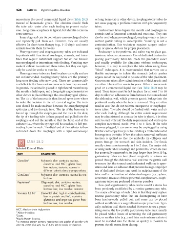
TABLE 28.2 passes through the stomach and skin incision. The fistula
usually closes spontaneously in 1 to 2 days. The major risk
Selected Enteral Diets of using such tubes is leakage and peritonitis, which are rare
but potentially catastrophic. In dogs larger than 20 to 25 kg,
DIET COMMENTS
gastrostomy tubes are best placed surgically or sutures are
Osmolite* Polymeric diet; contains taurine, passed through the abdominal wall and into the gastric wall
carnitine, and MCT; gluten free; to ensure that the stomach and abdominal wall stay in appo-
low lactose; isotonic, comes in sition and form an adhesion that prevents leakage. Improper
different caloric density preparations use of dedicated devices can result in malplacement of the
CliniCare † Polymeric diet; contains taurine but no tube and/or perforation of abdominal organs (e.g., spleen,
lactose omentum). Because of these potential complications, esoph-
Peptamen ‡ Oligomeric diet; contains taurine, agostomy tubes are preferred whenever possible.
carnitine, and MCT; gluten free; Low-profile gastrostomy tubes can be used if a stoma has
lactose free; low residue; isotonic been previously established by a routine gastrostomy tube.
Vivonex T.E.N. ‡ Elemental diet; high in carbohydrates, The major advantage of such tubes is that they may replace
¶
low in protein and fat ; contains routine gastrostomy tubes that are disintegrating or have
glutamine and arginine; gluten free; been inadvertently pulled out, and some can be placed
lactose free; low residue without anesthesia or a surgical/endoscopic procedure. Typi-
cally, sedation is all that is needed. However, to use a preex-
MCT, Medium-chain triglyceride. isting stoma, the low-profile gastrostomy tube must usually
*Abbott Nutrition. be placed within hours of removing the old gastrostomy
† Zoetis.
‡ Nestle Health Science. tube, or another tube (e.g., a red latex male urinary catheter)
¶ To increase protein content, reconstitute one packet of powder with must be inserted into the stoma as quickly as possible to
350 mL water plus 250 mL of 8.5% amino acids for injection. prevent the old stoma from closing.