
Page 539 - Small Animal Internal Medicine, 6th Edition
P. 539
CHAPTER 32 Disorders of the Peritoneum 511
VetBooks.ir
A B
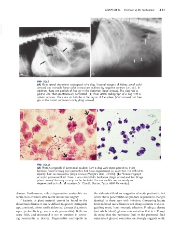
FIG 32.1
(A) Plain lateral abdominal radiograph of a dog. Visceral margins of kidney (small solid
arrows) and stomach (large solid arrows) are outlined by negative contrast (i.e., air). In
addition, there are pockets of free air in the abdomen (open arrows). This dog had a
gastric ulcer that spontaneously perforated. (B) Plain lateral radiograph of a dog with a
splenic abscess. There are air bubbles in the region of the spleen (short arrows) and free
gas in the dorsal peritoneal cavity (long arrows).
A B
FIG 32.2
(A) Photomicrograph of peritoneal exudate from a dog with septic peritonitis. Note
bacteria (small arrows) and neutrophils that have degenerated so much that it is difficult to
identify them as neutrophils (large arrows) (Wright’s stain; ×1000). (B) Photomicrograph
of septic peritoneal fluid. There is one intracellular bacterium (large arrow) and two things
(clear arrows) that may or may not be bacteria. The neutrophils are not nearly as
degenerated as in A. (A courtesy Dr. Claudia Barton, Texas A&M University.)
changes. Furthermore, mildly degenerative neutrophils are the abdominal fluid are suggestive of septic peritonitis, but
common in effusions after recent abdominal surgery. severe sterile pancreatitis can produce degenerative changes
If bacteria or plant material cannot be found in the identical to those seen with infection. Comparing lactate
abdominal effusion, it can be difficult to quickly distinguish levels in blood and effusion is not always accurate in distin-
septic peritonitis from sterile abdominal diseases that mimic guishing septic from nonseptic effusions. Finding a plasma
septic peritonitis (e.g., severe acute pancreatitis). Both can (not whole blood) glucose concentration that is > 38 mg/
cause SIRS, and ultrasound is not as sensitive in detect- dL more than the peritoneal fluid or the peritoneal fluid
ing pancreatitis as desired. Degenerative neutrophils in supernatant glucose concentration strongly suggests septic