
Page 563 - Small Animal Internal Medicine, 6th Edition
P. 563
CHAPTER 34 Diagnostic Tests for the Hepatobiliary and Pancreatic System 535
hypoxia and, in some cases, because of early disseminated enzymatic method for cats and dogs; 25 µmol/L with the
intravascular coagulation (DIC). Because increased pro- RIA method for dogs). Normal values for juvenile animals
VetBooks.ir duction and decreased excretion of bilirubin occur in dogs are similar to adult reference ranges. Abnormally high fasting
and/or postprandial SBA concentrations reflect a distur-
with severe hemolysis, serum bilirubin concentrations can
therefore be as high as 35 mg/dL. Icterus in cats with pure
the path of portal venous return to the liver and hepatocel-
hemolytic disease is an inconsistent finding and, if present, bance in hepatic secretion into the bile or at any point along
is mild. lular uptake.
Because almost all diseases associated with hyperbilirubi- The standard way to assess SBA concentration is outlined
nemia in cats and dogs are characterized by a mixture of in Box 34.3. Collective experience indicates that the likeli-
conjugated and unconjugated bilirubinemia, quantifying the hood of precipitating an episode of HE during this part of
two fractions by the use of the van den Bergh test achieves the test is extremely low, even in predisposed animals. After
little in regard to distinguishing primary hepatic or biliary the serum is harvested, the samples may be refrigerated for
disease from nonhepatobiliary disease in a clinical setting. several days or frozen almost indefinitely before assay. The
This lack of benefit in using the van den Bergh test may relate stability of the blood sample is one of the major advantages
to the time between onset of illness and examination, which over the much more labile serum ammonia test.
is usually at least several days. Under conditions of acute Studies of SBAs have confirmed their value in detecting
massive hemolysis, the total serum bilirubin concentration clinically relevant hepatobiliary disease requiring definitive
may initially consist primarily of the unconjugated form. As diagnostic testing in cats and dogs, especially in anicteric
hemolysis continues, the liver is able to take up and conju- animals with equivocal clinical signs and unexplained high
gate bilirubin, accounting for a combination of unconjugated liver enzyme activity. There continues to be controversy
and conjugated bilirubin. about whether a fasting or postprandial value alone is suffi-
Hyperbilirubinemia is attributed primarily to hemolysis cient or whether fasting and postprandial measurements are
when there is moderate to marked anemia with strong evi- required. If only one sample can be obtained, and the animal
dence of regeneration (except in the first 1-3 days, when the will eat or can tolerate being force-fed a small meal, the
response is less regenerative) and minimal changes in serum postprandial value is most useful to determine the presence
markers of cholestasis. Most dogs and cats with liver disease or absence, but not the type, of clinically relevant hepatobili-
are not severely anemic. ary disease in most cats and dogs. Current recommendations
state that for animals suspected of having acquired hepato-
Serum Bile Acid Concentration biliary disease, biopsy is needed when the postprandial SBA
The validation of rapid, technically simple methods for concentration using the enzymatic method in animals
serum bile acid (SBA) analysis in cats and dogs has provided without icterus exceeds 20 µmol/L in cats and 25 µmol/L in
a sensitive, variably specific test of hepatocellular function dogs. However, other researchers, particularly in the United
and the integrity of the enterohepatic portal circulation. Kingdom, have suggested that an SBA concentration of
Primary bile acids (e.g., cholic, chenodeoxycholic) are syn- between 20 and 40 µmol/L in dogs represents a gray area
thesized only in the liver, where they are conjugated with (Hall et al., 2005). Elevations in this range have been seen
various amino acids (primarily taurine) before secretion into
the bile. Bile is stored in the gallbladder, where it is concen-
trated until it is released into the duodenum under the influ-
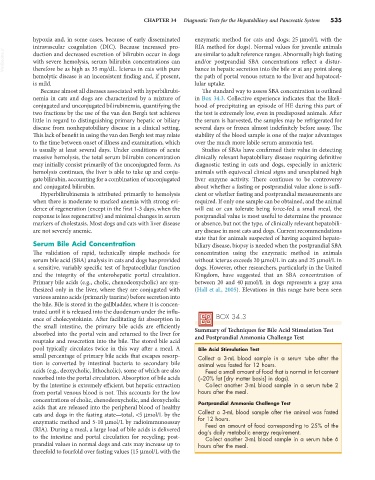
ence of cholecystokinin. After facilitating fat absorption in BOX 34.3
the small intestine, the primary bile acids are efficiently Summary of Techniques for Bile Acid Stimulation Test
absorbed into the portal vein and returned to the liver for and Postprandial Ammonia Challenge Test
reuptake and resecretion into the bile. The stored bile acid
pool typically circulates twice in this way after a meal. A Bile Acid Stimulation Test
small percentage of primary bile acids that escapes resorp- Collect a 3-mL blood sample in a serum tube after the
tion is converted by intestinal bacteria to secondary bile animal was fasted for 12 hours.
acids (e.g., deoxycholic, lithocholic), some of which are also Feed a small amount of food that is normal in fat content
resorbed into the portal circulation. Absorption of bile acids (≈20% fat [dry matter basis] in dogs).
by the intestine is extremely efficient, but hepatic extraction Collect another 3-mL blood sample in a serum tube 2
from portal venous blood is not. This accounts for the low hours after the meal.
concentrations of cholic, chenodeoxycholic, and deoxycholic Postprandial Ammonia Challenge Test
acids that are released into the peripheral blood of healthy
cats and dogs in the fasting state—total, <5 µmol/L by the Collect a 3-mL blood sample after the animal was fasted
for 12 hours.
enzymatic method and 5-10 µmol/L by radioimmunoassay Feed an amount of food corresponding to 25% of the
(RIA). During a meal, a large load of bile acids is delivered dog’s daily metabolic energy requirement.
to the intestine and portal circulation for recycling; post- Collect another 3-mL blood sample in a serum tube 6
prandial values in normal dogs and cats may increase up to hours after the meal.
threefold to fourfold over fasting values (15 µmol/L with the