
Page 613 - Small Animal Internal Medicine, 6th Edition
P. 613
CHAPTER 36 Hepatobiliary Diseases in the Dog 585
TABLE 36.1 BOX 36.1
VetBooks.ir Liver Diseases in Dogs SECONDARY Dog Breds Wih Reported Increased Risk of Chronic
Hepatitis*
PRIMARY
American and English Cocker Spaniels (worldwide,
Chronic hepatitis Steroid-induced hepatopathy males > females)
Copper storage disease Hepatic steatosis (lipidosis) Bedlington Terrier (worldwide, copper storage disease)
(secondary to diabetes Cairn Terrier (United Kingdom) †
mellitus or hypothyroidism) Dalmatian (United States, copper storage disease; United
†
Congenital portosystemic Congestion: heart failure or Kingdom, pathophysiology not reported )
shunt heartworm disease Doberman Pinschers (worldwide, some with copper
Drug- or toxin-induced Idiopathic vacuolar storage disease and some without; Scandinavian
reports suggest immune-mediated component, see text;
hepatopathy hepatopathy in Scottish females > males)
Terriers and others English Springer Spaniels (United Kingdom, Norway;
†
Reactive hepatitis (e.g., females > males)
secondary to pancreatitis, Great Dane (United Kingdom) †
inflammatory bowel Labrador Retrievers (worldwide; copper storage disease
disease) in United States and Holland; not copper-associated in
Metastatic neoplasia United Kingdom; females > males)
Samoyed (United Kingdom) †
Uncommon or Rare
West Highland White Terriers (worldwide; some copper-
Biliary tract disease, all Hepatocutaneous syndrome associated and some not)
types Standard Poodle (Anecdotal USA not copper associated).
Hepatic infections (see
text) *No reported sex ratio unless stated.
† Data for recently reported UK breeds from Bexfield NH, et al:
Portal vein hypoplasia,
microvascular dysplasia Breed, age, and gender distribution of dogs with chronic hepatitis
in the United Kingdom, Vet J 193:124, 2012.
Ductal plate abnormality Note previously reported hepatitis in Skye Terriers is now believed
Hepatic arteriovenous to be a congenital ductal plate abnormality (see later in chapter).
fistula
Acute fulminant hepatitis
(all causes)
Hepatic abscess
Primary neoplasia
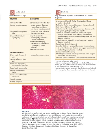
A B
FIG 36.1
(A) Histopathology of normal liver from a middle-aged Yorkshire Terrier. Note the normal
portal triad with hepatic portal vein, artery, and bile duct and hepatocytes arranged in
neat cords with sinusoids in between (white holes in bottom right are a sectioning artifact)
(H&E, ×200). (B) Histopathology of liver in a 3-year-old female English Springer Spaniel
with severe chronic hepatitis. There is marked distortion of the normal lobular structure
(compare to A), with inflammation, fibrosis, and hepatocyte vacuolation and necrosis.
There is also some ductular hyperplasia and disruption of the limiting plate (H&E, ×100).
(Courtesy Pathology Department, Veterinary Medicine, University of Cambridge,
Cambridge, England.)