
Page 617 - Small Animal Internal Medicine, 6th Edition
P. 617
CHAPTER 36 Hepatobiliary Diseases in the Dog 589
aspartate aminotransferase [AST]) and cholestatic enzymes
(alkaline phosphatase [ALP] and γ-glutamyltransferase
VetBooks.ir [GGT]) and evidence of decreased parenchymal liver func-
tion (low urea, low albumin, and sometimes high bilirubin
and bile acid concentrations). Persistent increases in ALT
levels are the most consistent finding in dogs with chronic
hepatitis but can also be found in other primary and second-
ary hepatopathies. A high ALP activity is much less specific
in dogs, particularly because there is a steroid-induced iso-
enzyme. Hepatocellular enzymes can become normal in end-
stage disease because of a lack of liver mass, but by that stage
function test results (e.g., ammonia and bile acid concentra-
tions) will be abnormal, and the dog may even be jaundiced.
Radiographic findings are nonspecific. Dogs with chronic
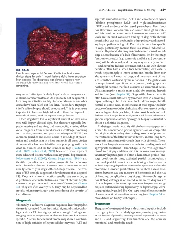
FIG 36.3 hepatitis often have a small liver (contrasting with cats, in
Liver from a 6-year-old Bearded Collie that had shown which hepatomegaly is more common), but the liver may
clinical signs for only 1 month before dying from end-stage also appear small in normal dogs, and the assessment of liver
liver disease. The diagnosis was chronic hepatitis with size is further confused by the variations in the gastric axis
macronodular cirrhosis and very little normal liver tissue in deep-chested dogs. If ascites is present, radiographs are
remaining. not helpful because the fluid obscures all abdominal detail.
Ultrasonography is much more useful for assessing hepatic
enzyme activities (particularly hepatocellular enzymes such architecture (see Chapter 34). Dogs with chronic hepatitis
as alanine aminotransferase [ALT]) should not be ignored. If often have a small, diffusely hyperechoic liver on ultrasonog-
liver enzyme activities are high for several months and other raphy, although the liver may look ultrasonographically
causes have been ruled out (see later, “Secondary Hepatopa- normal in some cases. In other cases it may appear nodular
thies”), a liver biopsy should be obtained. This is even more because of macronodular cirrhosis and/or concurrent benign
important in breeds at high risk and in those predisposed to nodular hyperplasia (see later). It is impossible to definitively
treatable diseases, such as copper storage disease. differentiate benign from malignant nodules on ultrasono-
Once dogs have lost a significant amount of liver mass, graphic appearance alone; cytology or biopsy is essential to
they will display clinical signs, but these are typically low- obtain a definitive diagnosis.
grade, waxing and waning, and nonspecific, making differ- End-stage chronic hepatitis with cirrhosis may appear very
ential diagnosis from other diseases a challenge. Vomiting similar to noncirrhotic portal hypertension or congenital
and diarrhea, anorexia, and polyuria-polydipsia (PU-PD) are ductal plate abnormality from a diagnostic standpoint, yet
common. Jaundice and ascites occur in some dogs at presen- the treatment of the latter is very different, and the long-term
tation and develop later in others but not in all cases. Ascites prognosis is much more favorable than with cirrhosis. There-
at presentation has been identified as a poor prognostic indi- fore a liver biopsy is necessary for a definitive diagnosis and
cator in humans and in two studies in dogs (Poldervaart appropriate treatment. Hemorrhage is the most significant
et al., 2009; Raffan et al., 2009) because it may represent risk of liver biopsy, and therefore it is the consensus amongst
more advanced disease with secondary portal hypertension. veterinary hepatologists to obtain a hemostasis profile (one-
Poldervaart et al. (2009); Gómez Selgas et al. (2014) also stage prothrombin time, activated partial thromboplastin
identified jaundice as a negative prognostic factor in dogs time, and platelet count) before obtaining a biopsy and to
with idiopathic chronic hepatitis. HE is uncommon and address any coagulopathies or thrombocytopenia before the
usually seen only in dogs with end-stage disease. The pres- procedure. However, publications fail to show a clear asso-
ence of HE strongly suggests the development of an acquired ciation between any one measure of hemostasis and the risk
PSS. Dogs with chronic hepatitis usually have some degree of bleeding complications postbiopsy. Fine-needle aspira-
of protein-calorie malnutrition as a result of chronic hepatic tion (FNA) cytology is of limited value in the diagnosis of
functional impairment and concurrent GI signs (see Chapter chronic hepatitis; the most representative biopsies are wedge
33). They are often overtly thin. They may be depressed but biopsies obtained during laparotomy or laparoscopy. Ultra-
are also often surprisingly alert considering the severity of sonographically guided Tru-Cut–type needle biopsies can be
their disease. of some benefit but are often misleading (see Chapter 34 for
more details on biopsy techniques).
Diagnosis
Ultimately, a definitive diagnosis requires a liver biopsy, but Treatment
disease is suspected from the clinical signs and clinicopatho- The goals of treatment of dogs with chronic hepatitis include
logic features. Clinical signs, clinicopathologic findings, and treating any identified underlying cause, slowing progression
imaging may be supportive of chronic hepatitis but are not of the disease if possible, treating clinical signs such as ascites
specific. A serum biochemical profile may show a combina- and HE, and supporting liver function and the animal’s
tion of high activities of hepatocellular enzymes (ALT and nutritional and metabolic needs.