
Page 641 - Small Animal Internal Medicine, 6th Edition
P. 641
CHAPTER 36 Hepatobiliary Diseases in the Dog 613
cytology analysis of the contents of a representative lesion at surgery, ranging in size from 2 to 5 cm in diameter; some
will distinguish an abscess from nodular hyperplasia, neo- dogs have a single nodule.
VetBooks.ir plasm (e.g., hemangiosarcoma), or granuloma. Ideally, mate- are identified only in liver biopsy specimens. The lesion con-
Micronodular changes occur much less frequently and
rial should be obtained for cytologic analysis and aerobic and
anaerobic bacterial cultures from a representative lesion
cytes with more mitotic figures and fewer binucleate cells
deep in the liver parenchyma to prevent abscess rupture and sists of increased numbers of normal to vacuolated hepato-
abdominal contamination. Abscess material should also be than expected in normal liver; components of normal lobular
obtained by this approach during surgery so that antibiotic architecture (e.g., portal tracts, central vein) remain. The
treatment can be initiated postoperatively. Ultrasound- adjacent parenchyma is compressed by growth of the nodules;
guided drainage of the abscess can also be used as treatment fibrosis, necrosis, inflammation, and bile ductule hyperplasia
in combination with appropriate antibiotics (see later). are absent. Because the prognosis for each of these nodular
Results of the preliminary clinicopathologic and radio- conditions is different and the margin of the lesion with
graphic evaluation should be scrutinized for evidence of adjacent hepatic tissue is important to establish a diagnosis,
previously noted comorbidities. a wedge biopsy is recommended. Needle specimens are likely
to be too small to confidently differentiate nodular hyperpla-
Treatment and Prognosis sia from primary hepatocellular carcinoma or adenoma. The
Treatment for liver abscesses consists of surgical removal of cause of this lesion is unknown; on the basis of the experi-
infected tissue, administration of appropriate antibiotics, mental development of nodular hyperplasia in rodent
supportive care, and resolution of underlying predisposing species, some have speculated a dietary role (low protein).
conditions. Infected liver tissue should be removed, if pos-
sible, and submitted for histopathologic examination and
bacterial culture if this was not done preoperatively. Fluid, NEOPLASIA
electrolyte, and acid–base abnormalities are addressed. Etiology
Administration of a combination of antibiotics with a gram-
negative and anaerobic spectrum is initiated until culture Primary hepatic neoplasms are rare in dogs, accounting for
and sensitivity test results are available. Because staphylo- fewer than 1.5% of all canine tumors. Unlike in cats, malig-
cocci and clostridia are the most common isolates, amoxicil- nant tumors are more common than benign tumors, and
lin (10–20 mg/kg IV q8h) combined with metronidazole metastatic tumors are 2.5 times more common than primary
(10 mg/kg PO q12h, or 7.5 mg/kg PO q12h for dogs with tumors in dogs. Metastases particularly arise from primary
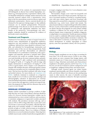
hepatic dysfunction) or clindamycin (10 mg/kg IV or PO neoplasms in the spleen, pancreas, and GI tract (Fig. 36.16);
q12h) is a good empiric choice. Surgery is not indicated for the liver can also be involved in systemic malignancies such
animals with multiple abscesses; ultrasound-guided centesis as lymphoma, malignant histiocytosis, and mastocytosis.
and abscess evacuation may be a reasonable adjunct to treat- Although certain chemical agents can induce hepatic
ment. This has been described combined with saline lavage neoplasms experimentally, and chronic hepatitis, steatohep-
and alcohol infusion. For more details, see Lemetayer et al. atitis, and chronic biliary tract disease are also predisposing
(2016). Antibiotic treatment is continued on a long-term causes in other species, the cause of naturally occurring
basis, usually for 6 to 8 weeks or until clinicopathologic and canine hepatic neoplasms is unknown. The types of primary
ultrasonographic indicators of abscessation are resolved.
From the limited information available about this rare condi-
tion, it seems that with aggressive medical and surgical treat-
ment, the prognosis for dogs with liver abscesses may not be
as poor as once thought.
NODULAR HYPERPLASIA
Hepatic nodular hyperplasia is a benign condition of older
dogs that does not cause clinical illness; clinicians should be
aware of it, however, because hyperplastic nodules may be
misinterpreted as a more serious condition, such as primary
or metastatic malignancy or regenerative nodules associated
with cirrhosis. The prevalence increases with age, and as
many as 70% to 100% of dogs older than 14 years have some
microscopic or macroscopic hyperplasia. Affected dogs have
high serum ALP activities (usually a 2.5-fold elevation but
possibly as high as 14-fold), which prompts an investigation
for hyperadrenocorticism. There is no evidence of hepatic FIG 36.16
dysfunction on serum biochemical analysis. Many dogs have Gross appearance of liver postmortem from a 2-year-old
multiple macroscopic nodules found ultrasonographically or male Siberian Husky with a metastatic carcinoma.