
Page 645 - Small Animal Internal Medicine, 6th Edition
P. 645
CHAPTER 36 Hepatobiliary Diseases in the Dog 617
VetBooks.ir
A B
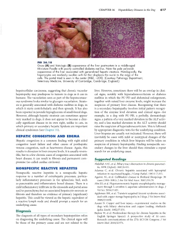
FIG 36.18
Gross (A) and histologic (B) appearance of the liver postmortem in a middle-aged
Miniature Poodle with poorly controlled diabetes mellitus. Note the pale yellowish
appearance of the liver associated with generalized hepatic steatosis. Histologically, the
hepatocytes are markedly swollen with fat that displaces the nuclei to the edge of the
cells. The portal triad is seen in the center (H&E, ×200). (Courtesy Pathology Department,
Veterinary Medicine, University of Cambridge, Cambridge, England.)
hepatocellular carcinoma, suggesting that chronic vacuolar liver. However, sometimes there will be an overlap in clini-
hepatopathy may predispose to tumors in dogs as it can in cal signs, notably with hyperadrenocorticism or diabetes
humans. The vacuolation seen as part of the hepatocutane- mellitus in which the PU-PD and abdominal enlargement,
ous syndrome looks similar to glycogen vacuolation. Steato- together with raised liver enzyme levels, might increase the
sis is generally associated with diabetes mellitus in dogs, in suspicion of primary liver disease. Recognizing that there
which it starts centrilobularly and then spreads. It has also is a secondary hepatopathy involves initial pattern recogni-
been reported in juvenile hypoglycemia of small-breed dogs. tion of the enzyme level elevation and clinical signs—for
However, although hepatic steatosis can sometimes appear example, in a dog with PU-PD, a potbelly, dermatologic
very marked in dogs, it does not appear to become a clini- signs, a pattern of a very marked elevation in the ALP activ-
cally significant disease in its own right, unlike in cats, in ity, and a less marked elevation in the ALT activity should
which primary or secondary hepatic lipidosis are important raise the suspicion of hyperadrenocorticism. This is followed
clinical syndromes (see Chapter 35). by appropriate diagnostic tests for the underlying condition.
Liver biopsies are usually not indicated. However, there will
HEPATIC CONGESTION AND EDEMA inevitably be cases with mild or nontypical changes of the
Hepatic congestion is a common finding with right-sided primary condition in which liver biopsies will be taken on
congestive heart failure and other causes of posthepatic suspicion of primary hepatopathy. Finding nonspecific sec-
venous congestion, such as heartworm disease. Again, this ondary changes in the liver should then stimulate a repeat
results in elevation in liver enzyme levels. It is usually revers- search for an underlying cause.
ible, but in a few chronic cases of congestion associated with
heart disease, it can result in fibrosis and permanent com- Suggested Readings
promise (so-called cardiac cirrhosis).
Abdallah AAL, et al. Biliary tract obstruction in chronic pancreati-
tis. HPB (Oxford). 2007;9:421.
NONSPECIFIC REACTIVE HEPATITIS Adamus C, et al. Chronic hepatitis associated with leptospiral
Nonspecific reactive hepatitis is a nonspecific hepatic infection in vaccinated beagles. J Comp Pathol. 1997;117:311.
response to a number of extrahepatic processes, particu- Aguirre AL, et al. Gallbladder disease in Shetland Sheepdogs: 38
larly inflammatory processes in the splanchnic bed, such cases (1995-2005). J Am Vet Med Assoc. 2007;231:79.
as pancreatitis and inflammatory bowel disease. There is a Ahn JO, et al. Hyperammonemic hepatic encephalopathy manage-
mild inflammatory infiltrate in the sinusoids and portal areas ment through L-ornithin-L-aspartate administration in dogs. J
and/or parenchyma but no associated hepatocyte necrosis or Vet Sci. 2016;17:431.
fibrosis and therefore no evidence of primary (significant) Appleman EH, et al. Transient acquired fanconi syndrome associ-
hepatitis. This could be viewed as the hepatic equivalent of ated with copper storage hepatopathy in 3 dogs. J Vet Intern Med.
2008;22:1038.
a reactive lymph node and should prompt a search for an Azumi N. Copper and liver injury—experimental studies on the
underlying cause. dogs with biliary obstruction and copper loading. Hokkaido
Diagnosis Igaku Zasshi. 1982;57:331.
Bayton W, et al. Prednisolone therapy for chronic hepatitis in the
The diagnosis of all types of secondary hepatopathies relies English Springer Spaniel: A prospective study of 14 cases.
on diagnosing the underlying cause. The clinical signs will Research communications of the 27th ECVIM Congress. J Vet
be those of the primary cause and are not related to the Intern Med. 2018;32:574.