
Page 1001 - Adams and Stashak's Lameness in Horses, 7th Edition
P. 1001
Occupational‐Related Lameness Conditions 967
Abaxial fragments are not articular and surgical removal
should not be attempted.
VetBooks.ir straightforward; however, special mention should be
Fractures of the first phalanx occur and are generally
made of incomplete sagittal fractures because they are
common in the Standardbred and can represent a diag
nostic challenge. Marked lameness after a race subsides
quickly but recurs with return to work. Heat, swelling,
and joint distention may be absent or minimal. This sce
nario, coupled with a positive response to fetlock flex
ion, should arouse suspicion. Radiographs prior to
diagnostic anesthesia are warranted because propaga
tion of the fracture can occur. Short fractures may not be
radiographically visible until bone resorption occurs
3–6 weeks following fracture. Diagnosis is facilitated by
observation of linear lucency in the proximal sagittal
groove on the AP view and dorsal periosteal remodeling,
which is best seen on the lateral to medial projection.
Sclerosis near the midsagittal groove often precedes
fracture, and nuclear scintigraphy is likely to demon
strate the lesion much earlier. Until an accurate diagno
sis can be made, these cases should be managed
conservatively in a stall to prevent catastrophic injury.
Short fractures may be treated with confinement alone,
whereas longer, propagating, or nonhealing fractures
benefit from internal fixation. Healing occurs in 4–6
months, and the prognosis for racing is good, provided
significant degenerative changes do not occur. 8
Cyclic bone fatigue occurs in the Standardbred when
bone adapts to the miles of low‐speed jogging one way
of the track and is then unable to endure the intense
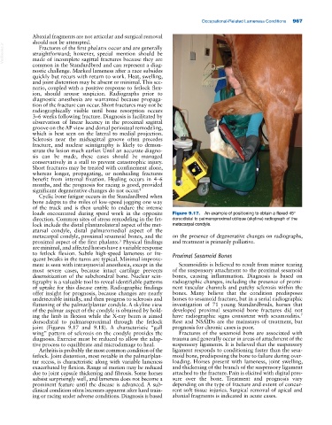
loads encountered during speed work in the opposite Figure 9.17. An example of positioning to obtain a flexed 45°
direction. Common sites of stress remodeling in the fet dorsodistal to palmaroproximal oblique (skyline) radiograph of the
lock include the distal plantarolateral aspect of the met metacarpal condyle.
atarsal condyle, distal palmaromedial aspect of the
metacarpal condyle, proximal sesamoid bones, and the on the presence of degenerative changes on radiographs,
proximal aspect of the first phalanx. Physical findings and treatment is primarily palliative.
3
are minimal, and affected horses have a variable response
to fetlock flexion. Subtle high‐speed lameness or fre Proximal Sesamoid Bones
quent breaks in the turns are typical. Minimal improve
ment is seen with intrasynovial anesthesia, except in the Sesamoiditis is believed to result from minor tearing
most severe cases, because intact cartilage prevents of the suspensory attachment to the proximal sesamoid
desensitization of the subchondral bone. Nuclear scin bones, causing inflammation. Diagnosis is based on
tigraphy is a valuable tool to reveal identifiable patterns radiographic changes, including the presence of promi
of uptake for this disease entity. Radiographic findings nent vascular channels and patchy sclerosis within the
offer insight for prognosis, because changes are nearly bones. Many believe that the condition predisposes
undetectable initially, and then progress to sclerosis and horses to sesamoid fracture, but in a serial radiographic
flattening of the palmar/plantar condyle. A skyline view investigation of 71 young Standardbreds, horses that
of the palmar aspect of the condyle is obtained by hold developed proximal sesamoid bone fractures did not
ing the limb in flexion while the X‐ray beam is aimed have radiographic signs consistent with sesamoiditis.
7
dorsodistal to palmaroproximal through the fetlock Rest and NSAIDs are the mainstays of treatment, but
joint (Figures 9.17 and 9.18). A characteristic “gull prognosis for chronic cases is poor.
wing” pattern of sclerosis on the condyle provides the Fractures of the sesamoid bone are associated with
diagnosis. Exercise must be reduced to allow the adap trauma and generally occur in areas of attachment of the
tive process to equilibrate and microdamage to heal. suspensory ligaments. It is believed that the suspensory
Arthritis is probably the most common condition of the ligament responds to conditioning faster than the sesa
fetlock. Joint distention, most notable in the palmar/plan moid bone, predisposing the bone to failure during over
tar recess, is characteristic along with variable lameness loading. Horses present with lameness, joint swelling,
exacerbated by flexion. Range of motion may be reduced and thickening of the branch of the suspensory ligament
due to joint capsule thickening and fibrosis. Some horses attached to the fracture. Pain is elicited with digital pres
subsist surprisingly well, and lameness does not become a sure over the bone. Treatment and prognosis vary
prominent feature until the disease is advanced. A sub depending on the type of fracture and extent of concur
clinical condition often becomes apparent after hard train rent soft tissue injuries. Surgical removal of apical and
ing or racing under adverse conditions. Diagnosis is based abaxial fragments is indicated in acute cases.