
Page 1003 - Adams and Stashak's Lameness in Horses, 7th Edition
P. 1003
Occupational‐Related Lameness Conditions 969
lameness in 30% of horses, with severity directly related may require regular injections to remain competitive.
9
to the degree of third carpal bone sclerosis. Increased Flat shoes with square toes ease break‐over, and the
VetBooks.ir necrosis and predispose to further injury (Figure 9.19). shear strain.
removal of all shoe additives (toe grabs) helps to reduce
bone density and loss of vascular channels cause ischemic
19
Distention of the tarsocrural joint is usually associ
Lucency of C3 is also associated with lameness,
14,17,19
and lesions in bone and cartilage are usually identified ated with osteochondrosis (OCD) that is prevalent in
at surgery. Etiopathogenesis is similar to that seen in the Standardbred. Frequency and distribution of OCD
14
the fetlock and requires similar management. lesions may differ between trotters and pacers, and
11
Incidence of carpal slab fractures is highest in 2‐year‐ early removal of most fragments allows horses to per
olds, with frontal plane fractures of the radial facet of form equivalently to unaffected counterparts. 11
the third carpal bone the most common configuration. Fractures of the central or third tarsal bone are occa
14
Distribution is nearly equal between the right and left sionally seen in the racing Standardbred. Lameness can
limbs. Horses are lame and resentful of flexion, often be severe, but other external signs are often limited and
leaning back or rearing to avoid it. Diagnosis is con do not specifically incriminate the distal tarsus. Both
firmed using radiography; the tangential view (skyline) conservative treatment and internal fixation have pro
of the third carpal bone is the most valuable. Fractures vided a fair prognosis for return to racing, but arthritis
heal with conservative management, but surgery limits remains an inevitable sequellae. 13,20
the severity of joint deterioration and provides the best Curbs frequently develop in 2‐year‐olds as speed
chance to return to racing. Up to 77% of Standardbreds training begins. In addition to swelling in the long plan
return to racing following slab fracture, but earnings tar ligament, injury to the digital flexor tendons, collat
may be decreased. 18 eral ligaments, and numerous other structures in this
15
region can lead to the appearance of curb. Cryotherapy
(freeze firing) is often employed for persistently painful
Tarsus
curbs.
Young Standardbreds with uneven gaits or those
subjected to miles of jogging quickly develop tarsitis. Stifle
Affected horses exhibit a shortened stride and may stab
the toe laterally as they land, causing uneven wear to The stifle is frequently implicated as a source of lame
the shoe. Disease is most frequent in the tarsometatar ness in the Standardbred, but a thorough work‐up is
sal joint but can occur solely in the distal intertarsal rarely performed at the track, and the true incidence of
joint; therefore, these joints should be blocked (and lameness attributable to this region is unknown. Usually,
treated) separately. Nuclear scintigraphy of the lameness blocks out lower in the limb. Horses with stifle
Standardbred with distal tarsitis reveals increased radi lameness tend to carry the affected limb wide and land
opharmaceutical uptake (IRU) in the dorsolateral on the toe. Trotters may develop a bunny hop gait, par
region of the joints, in contrast to a medial location ticularly if affected bilaterally.
seen in most other sport horses. Radiographic findings Standardbreds with so‐called loose stifles frequently
4
correlate poorly with disease severity, and they are have quadriceps muscle soreness that makes them
frequently negative in 2‐ and 3‐year‐olds despite a resemble horses with intermittent upward fixation of
response to diagnostic anesthesia. Intra‐articular use of the patella. They develop an abnormal cranial phase of
HA and corticosteroids is beneficial, and older horses the stride as the patella momentarily “catches” before the
limb is brought to the ground. Generally, this is a fitness
issue that is cured by trotting up and down hills or
working in deep footing to hasten conditioning.
Nevertheless, many veterinarians advocate internal blis
ters over the medial patellar ligament to induce scarring
and decrease laxity. Medial patellar desmotomy should
be reserved for cases of true upward fixation because
fragmentation of the distal end of the patella has been
reported following this procedure in normal horses. 12
Stifle swelling should be a red flag whenever it is
found. Marked femoropatellar effusion with minor
lameness is characteristic of OCD and should prompt
radiographic investigation. The postoperative progno
5
sis is favorable in the Standardbred, as long as lesions
are not extensive. Medial femorotibial effusion is a more
subtle but ominous sign, often indicating advanced
arthritis or injury to the intra‐articular support struc
tures of the stifle. These injuries usually end a racing
career.
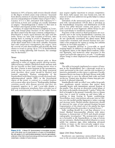
Figure 9.19. A flexed 35° dorsoproximal to dorsodistal (skyline)
view of the distal row of carpal bones in a 3‐year‐old Standardbred Upper Limb Stress Fractures
filly with grade 2 of 5 right forelimb lameness. Note the generalized
sclerosis of the third carpal bone along with focal lucent areas in the Racehorses can experience stress‐related bone dis
radial facet, indicative of bone necrosis. ease in the proximal appendicular skeleton, including