
Page 1189 - Adams and Stashak's Lameness in Horses, 7th Edition
P. 1189
Miscellaneous Musculoskeletal Conditions 1155
Traumatic wounds that involve a bony surface or resonance imaging (MRI) and scintigraphy are the
chronic synovial sepsis are most likely to result in osteo modalities of choice for suspected cases of human osteo
VetBooks.ir matory or infective process of the cortex and marrow earlier diagnosis in horses. Ultrasonography also can
myelitis and septic arthritis and may also provide for an
myelitis or osteitis. Osteomyelitis refers to the inflam
20
be effective in helping to diagnose osteomyelitis by
components of bone. If only cortical bone is infected,
17
17
it is classified as osteitis. Penetrating wound tracts that imaging fluid pockets in and around the infected bone. 41
lead to bone should be treated aggressively. The presence
of osteomyelitis/osteitis is significantly associated with Treatment
nonsurvival in cases with synovial sepsis (Figure 12.20). 57, 61
Radiography is commonly used to diagnose osteomyeli Acute wounds involving synovial structures, includ
tis/osteitis, but unfortunately, 30–50% of bone deminer ing joints, bursae, and tendon sheaths, often directly
alization must occur before it can be observed on plain introduce bacteria and other contaminants into the syn
radiographs. This may require 14–21 days, resulting in ovial space. The clinician’s primary concern is removing
very low sensitivity and a delayed diagnosis. magnetic the bacteria from the space before infection can become
established. Early recognition and treatment of a syno
vial penetrating wound are imperative to reduce the risk
of developing septic arthritis. One study reported that
horses treated within 24 hours had a lower risk of devel
oping septic arthritis compared with those treated after
24 hours. Wereszka et al. found that horses with sep
16
58
tic tenosynovitis were significantly more likely to sur
vive if treated during the first day after clinical signs of
synovial infection were observed than horses that did
not receive treatment within 10 days after clinical signs
of infection appeared. However, more recent publica
tions reported that the duration of time until treatment
may not affect survival 33,57 or the ability to return to
57
athletic function. Logically, it would still be advised
that the quicker or earlier these cases are treated, the
better the success (Figure 12.21).
Any wound that communicates with a synovial structure
or bone should be considered contaminated. Therefore,
early treatment with antimicrobials may help decrease the
progression to synovial sepsis and/or osteomyelitis/osteitis.
Antimicrobials can be administered parenterally, region
ally, locally, or a combination of all. Additional therapies
for preventing infection in a synovial cavity include some
form of synovial lavage, drainage or endoscopic explora
tion, and wound debridement with or without closure.
Small wounds with minimal contamination can be man
aged conservatively, but the clinical response should be
monitored closely, and any deterioration should be met
with more aggressive treatment.
Systemic antibiotics can be initiated for any infected
wound, regardless of the structures, to provide a broad
spectrum of activity against common microbes. These
antibiotics are maintained until sensitivity of the cul
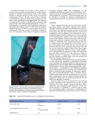
Figure 12.19. This traumatic wound was associated with an tured organism is obtained or concerns about sepsis
open comminuted fracture of the fourth metatarsus. Wound have diminished. Most commonly, a combination of
debridement and lavage are important to prevent osteomyelitis in penicillin (22,000 IU/kg every 6 hours IV, every 12 hours
these horses. Source: Courtesy of Dr. Gary Baxter.
Table 12.2. Synovial fluid parameters used to aid diagnosis of synovial sepsis.
Fluid analysis parameter Normal joint Traumatic synovitis Septic synovitis
Total protein (g/L) 18 ± 3 20–40 >40
Less than 20
Total nucleated cell count (× 10 /L) <1.0 <10.0 >30.0
9
<3.5 (tendon sheath)
Neutrophils (%) <10 <10 >80