
Page 1192 - Adams and Stashak's Lameness in Horses, 7th Edition
P. 1192
1158 Chapter 12
screw with a Luer lock head is placed in the drilled and
tapped hole. Alternatively, the end of a catheter extension
VetBooks.ir process is very similar: a tourniquet is placed proximal to
set fits securely into the drilled hole. The remainder of the
the third metacarpal/metatarsal bone. The infusate, of
similar volume and concentration as with IVRLP, is
slowly injected avoiding excess pressure. The tourniquet
should be left in place for 20–30 minutes following the
injection. Intraosseous perfusion may be favored over
IVRLP in cases that lack easy venous access or the pres
ence of osteomyelitis. Data suggests osteomyelitic lesions
may respond better to intraosseous perfusion. 29
Antibiotic‐Impregnated Materials
Antibiotics can be effectively delivered by slow
release from impregnated materials. These products are
left in situ to release the drug over an extended period
of time, resulting in high local tissue concentrations.
The most common depot material used for local antibi
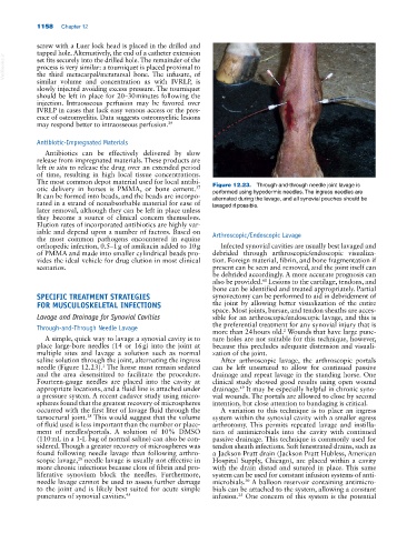
otic delivery in horses is PMMA, or bone cement. Figure 12.23. Through‐and‐through needle joint lavage is
37
It can be formed into beads, and the beads are incorpo performed using hypodermic needles. The ingress needles are
alternated during the lavage, and all synovial pouches should be
rated in a strand of nonabsorbable material for ease of lavaged if possible.
later removal, although they can be left in place unless
they become a source of clinical concern themselves.
Elution rates of incorporated antibiotics are highly var
iable and depend upon a number of factors. Based on Arthroscopic/Endoscopic Lavage
the most common pathogens encountered in equine
orthopedic infection, 0.5–1 g of amikacin added to 10 g Infected synovial cavities are usually best lavaged and
of PMMA and made into smaller cylindrical beads pro debrided through arthroscopic/endoscopic visualiza
vides the ideal vehicle for drug elution in most clinical tion. Foreign material, fibrin, and bone fragmentation if
scenarios. present can be seen and removed, and the joint itself can
be debrided accordingly. A more accurate prognosis can
also be provided. Lesions to the cartilage, tendons, and
61
bone can be identified and treated appropriately. Partial
SPECIFIC TREATMENT STRATEGIES synovectomy can be performed to aid in debridement of
FOR MUSCULOSKELETAL INFECTIONS the joint by allowing better visualization of the entire
space. Most joints, bursae, and tendon sheaths are acces
Lavage and Drainage for Synovial Cavities sible for an arthroscopic/endoscopic lavage, and this is
Through‐and‐Through Needle Lavage the preferential treatment for any synovial injury that is
2
more than 24 hours old. Wounds that have large punc
A simple, quick way to lavage a synovial cavity is to ture holes are not suitable for this technique, however,
place large‐bore needles (14 or 16 g) into the joint at because this precludes adequate distension and visuali
multiple sites and lavage a solution such as normal zation of the joint.
saline solution through the joint, alternating the ingress After arthroscopic lavage, the arthroscopic portals
1
needle (Figure 12.23). The horse must remain sedated can be left unsutured to allow for continued passive
and the area desensitized to facilitate the procedure. drainage and repeat lavage in the standing horse. One
Fourteen‐gauge needles are placed into the cavity at clinical study showed good results using open wound
appropriate locations, and a fluid line is attached under drainage. It may be especially helpful in chronic syno
49
a pressure system. A recent cadaver study using micro vial wounds. The portals are allowed to close by second
spheres found that the greatest recovery of microspheres intention, but close attention to bandaging is critical.
occurred with the first liter of lavage fluid through the A variation to this technique is to place an ingress
tarsocrural joint. This would suggest that the volume system within the synovial cavity with a smaller egress
28
of fluid used is less important than the number or place arthrotomy. This permits repeated lavage and instilla
ment of needles/portals. A solution of 10% DMSO tion of antimicrobials into the cavity with continued
(110 mL in a 1‐L bag of normal saline) can also be con passive drainage. This technique is commonly used for
sidered. Though a greater recovery of microspheres was tendon sheath infections. Soft fenestrated drains, such as
found following needle lavage than following arthro a Jackson Pratt drain (Jackson Pratt Hubless, American
scopic lavage, needle lavage is usually not effective in Hospital Supply, Chicago), are placed within a cavity
28
more chronic infections because clots of fibrin and pro with the drain distad and sutured in place. This same
liferative synovium block the needles. Furthermore, system can be used for constant infusion systems of anti
needle lavage cannot be used to assess further damage microbials. A balloon reservoir containing antimicro
30
to the joint and is likely best suited for acute simple bials can be attached to the system, allowing a constant
punctures of synovial cavities. 45 infusion. One concern of this system is the potential
25