
Page 166 - Adams and Stashak's Lameness in Horses, 7th Edition
P. 166
132 Chapter 2
Hip hike
VetBooks.ir
Hip dip
A B
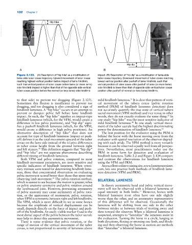
Figure 2.123. (A) Description of “hip hike” as a modification of impact. (B) Description of “hip dip” as a modification of lame‐side
lame‐side tuber coxae trajectory. Upward movement of tuber coxae tuber coxae trajectory. Downward movement of tuber coxae reaching
reaching highest vertical position before impact of lame hindlimb, lowest vertical position after pushoff of lame hindlimb, such that
such that vertical position of tuber coxae before lame (or more lame) vertical position of tuber coxae after pushoff of lame (or more lame)
side hindlimb impact is higher than that of the opposite side vertical side hindlimb is lower than that of opposite side vertical tuber coxae
tuber coxae position before the normal (or less lame) side hindlimb position after pushoff of normal (or less lame) hindlimb.
to that side) to prevent toe dragging (Figure 2.123). mild hindlimb lameness. It is clear that pattern of verti
51
Sometimes this flexion is insufficient to prevent toe cal movement of the tubera coxae (pelvic rotation
dragging, and toe dragging is also considered a sign of method [PRM] of hindlimb lameness detection) does
hindlimb lameness. A “hip hike” occurs in an attempt to not accurately quantify the true state of vertical tubera
prevent or dampen pelvic fall before lame hindlimb sacral movement (VPM method) and vice versa; in other
impact. As such, the “hip hike” signifies an impact‐type words, they do not exactly evaluate the same thing. In
51
hindlimb lameness (which, for the VPM, would create a one study “hip hike” was the most sensitive indicator of
difference in low pelvic positions), and “hip dip” signi mild hindlimb lameness. In one study vertical move
40
fies a pushoff hindlimb lameness (which, for the VPM, ment of the tuber sacrale had the highest discriminating
would create a difference in high pelvic positions). An power for determination of hindlimb lameness. 15
alternative description of “hip hike” that does not The best position for the evaluator using the PRM is
account for type of hindlimb lameness (impact or push behind the horse with the horse moving away from the
off) defines it as the total movement upward of the tuber evaluator with spatial resolution of the observer degrad
coxae on the lame side instead of the relative differences ing with each stride. The VPM method is more versatile
in tuber coxae height from the ground between right because it can be observed equally well from all perspec
and left stances. This definition suggests that “hip dip” tives. Nevertheless, most practitioners today use the
40
and “hip hike” are not separate phenomena describing PRM in some form for detection and evaluation of
different timing of hindlimb lameness. hindlimb lameness. Table 2.2 and Figure 2.124 compare
Both VPM and pelvic rotation, compared to most and contrast the observations for hindlimb lameness
hindlimb movement parameters, are more sensitive and using the VPM and PRM.
specific indicators of hindlimb lameness. When veteri An excellent online training site, www.lamenesstrainer.
nary trainees were tested for determining hindlimb lame com, lets one practice both methods of hindlimb lame
ness, those that concentrated observation on evaluating ness detection (VPM and PRM).
pelvic movement scored better than those that spent time
50
observing limb movement. The pelvis rotation method, BILATERAL LAMENESS
which is easiest to see because the motion is greater, relies
on pelvic anatomy symmetry and pelvic rotation around In theory asymmetric head and pelvic vertical move
the lumbosacral joint. However, preexisting asymmetry ment will not be observed with a bilateral lameness of
of pelvic anatomy may cause asymmetry of pelvic rota equal intensity in both limbs. However, horses with
11
tion, or the appearance of asymmetric pelvic rotation, bilateral lameness most often have one side slightly
when VPM is symmetric between right and left hindlimbs. worse than the other, and an asymmetry representative
The VPM, which is more difficult to see in some horses of this difference will be observed. Occasionally the
because the amplitude of vertical displacement can be lameness will be shifting from one side to the other
small, depends directly on the force of impact and push between strides or between trials of trotting away and
off of the hindlimbs. An easily visible marker fixed to the then toward the observer. When a bilateral lameness is
most dorsal aspect of the pelvis between the tuber sacrale suspected, attempts to “lateralize” the lameness assist in
may help to detect this asymmetric movement. the evaluation. Turning the horse in a circle, lunging in
There is some evidence that pelvic rotation, or the both directions, flexing or stressing one limb, and block
range of motion of the vertical movement of the tuber ing and then observing the horse in motion are methods
coxae, is not proportional to severity of lameness above that “lateralize” a bilateral lameness.