
Page 274 - Adams and Stashak's Lameness in Horses, 7th Edition
P. 274
VetBooks.ir
14
1
13
2
12
3
a b 4
11
5
10
9
6
8 7
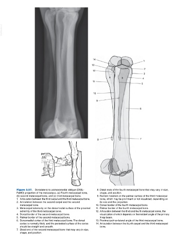
Figure 3.57. Dorsolateral to palmaromedial oblique (D55L‐ 8. Distal ends of the fourth metacarpal bone that may vary in size,
PaMO) projection of the metacarpus. (a) Fourth metacarpal bone, shape, and position.
(b) second metacarpal bone, and (c) third metacarpal bone. 9. Nutrient foramen on the palmar surface of the third metacarpal
1. Articulation between the third carpal and the third metacarpal bone. bone, which may be prominent or not visualized, depending on
2. Articulation between the second carpal and the second its size and the projection.
metacarpal bone. 10. Dorsal border of the fourth metacarpal bone.
3. Metacarpal tuberosity on the dorsomedial surface of the proximal 11. Palmar border of the fourth metacarpal bone.
extremity of the third metacarpal bone. 12. Articulation between the third and fourth metacarpal bones, the
4. Dorsal border of the second metacarpal bone. visualization of which depends on the incident angle of the primary
5. Palmar border of the second metacarpal bone. X‐ray beam.
6. Dorsomedial cortex of the third metacarpal bone. The dorsal 13. Proximal‐palmar‐lateral angle of the third metacarpal bone.
cortex is normally thick, and the periosteal surface of the cortex 14. Articulation between the fourth carpal and the third metacarpal
should be straight and smooth. bone.
7. Distal end of the second metacarpal bone that may vary in size,
shape, and position.