
Page 278 - Adams and Stashak's Lameness in Horses, 7th Edition
P. 278
VetBooks.ir
28
27
1
26
2
a 3
25 4
24 b 5
23
6
22 7
8
21 9
10
11
20 12
19
c
18
17
16
15
14 13
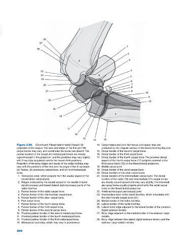
Figure 3.59. (Continued) Flexed lateromedial (flexed LM) 14. Carpometacarpal joint. Numerous joint space lines are
projection of the carpus. The size and shape of the first and fifth produced by the irregular contour of the bones forming this joint.
carpal bones may vary, and sometimes the bones are absent. The 15. Dorsal border of the second carpal bone.
palmar borders of the carpal and metacarpal bones are closely 16. Dorsal border of the third carpal bone.
superimposed in this projection, and the positions may vary slightly 17. Dorsal border of the fourth carpal bone. The proximal dorsal
with X‐ray tube angulation and/or the horse’s limb positions. aspect of the fourth carpal bone (17) projects proximal to the
Projection of the bony ridges and facets of the radial trochlea may third carpal bone (16) on the flexed lateral projection.
vary with the positions of the limb and the angle of the X‐ray beam. 18. Middle carpal joint.
(a) Radius, (b) accessory carpal bone, and (c) third metacarpal 19. Dorsal border of the radial carpal bone.
bone. 20. Dorsal borders of the ulnar carpal bone.
1. Transverse crest, which projects from the caudal aspect of the 21. Dorsal borders of the intermediate carpal bone. The dorsal
closed distal radial physis. borders of the radial (19) and intermediate (21) carpal bones
2. Ridges produced by the caudal aspect of the medial (medial are closely superimposed and may vary slightly. The intermedi
styloid process) and lateral (lateral styloid process) parts of the ate carpal bone usually projects proximal to the radial carpal
radial trochlea. bone on the flexed lateral projection.
3. Palmar border of the radial carpal bone. 22. Antebrachiocarpal (radiocarpal) joint.
4. Palmar border of the intermediate carpal bone. 23. Intermediate facet of the radial trochlea, which articulates with
5. Palmar border of the ulnar carpal bone. the intermediate carpal bone (21).
6. First carpal bone. 24. Medial border of the radial trochlea.
7. Palmar border of the fourth carpal bone. 25. Lateral border of the radial trochlea.
8. Palmar border of the third carpal bone. 26. Lateral bony ridge adjacent to the lateral border of the common
9. Palmar border of the second carpal bone. digital extensor tendon.
10. Proximal palmar border of the second metacarpal bone. 27. Bony ridge adjacent to the medial border of the extensor carpi
11. Proximal palmar border of the fourth metacarpal bone. radialis.
12. Proximal palmar border of the third metacarpal bone. 28. Bony ridge between the lateral digital extensor tendon and the
13. Metacarpal tuberosity, which may vary in prominence. extensor carpi radialis tendon.
244