
Page 288 - Adams and Stashak's Lameness in Horses, 7th Edition
P. 288
VetBooks.ir
11
1
10
9
2
8
7
6
6
5
4 6 3
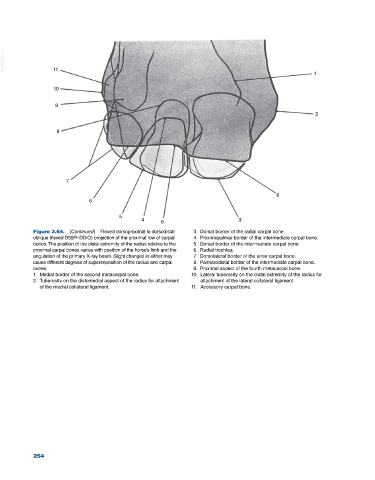
Figure 3.64. (Continued) Flexed dorsoproximal to dorsodistal 3. Dorsal border of the radial carpal bone.
oblique (flexed D55Pr‐DDiO) projection of the proximal row of carpal 4. Proximopalmar border of the intermediate carpal bone.
bones. The position of the distal extremity of the radius relative to the 5. Dorsal border of the intermediate carpal bone.
proximal carpal bones varies with position of the horse’s limb and the 6. Radial trochlea.
angulation of the primary X‐ray beam. Slight changes in either may 7. Dorsolateral border of the ulnar carpal bone.
cause different degrees of superimposition of the radius and carpal 8. Palmarodistal border of the intermediate carpal bone.
bones. 9. Proximal aspect of the fourth metacarpal bone.
1. Medial border of the second metacarpal bone. 10. Lateral tuberosity on the distal extremity of the radius for
2. Tuberosity on the distomedial aspect of the radius for attachment attachment of the lateral collateral ligament.
of the medial collateral ligament. 11. Accessory carpal bone.
254