
Page 292 - Adams and Stashak's Lameness in Horses, 7th Edition
P. 292
VetBooks.ir
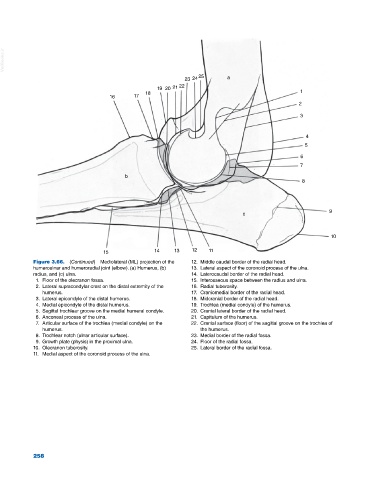
23 24 25 a
19 20 21 22
18 1
16 17
2
3
4
5
6
7
b
8
9
c
10
15 14 13 12 11
Figure 3.66. (Continued) Mediolateral (ML) projection of the 12. Middle caudal border of the radial head.
humeroulnar and humeroradial joint (elbow). (a) Humerus, (b) 13. Lateral aspect of the coronoid process of the ulna.
radius, and (c) ulna. 14. Laterocaudal border of the radial head.
1. Floor of the olecranon fossa. 15. Interosseous space between the radius and ulna.
2. Lateral supracondylar crest on the distal extremity of the 16. Radial tuberosity.
humerus. 17. Craniomedial border of the radial head.
3. Lateral epicondyle of the distal humerus. 18. Midcranial border of the radial head.
4. Medial epicondyle of the distal humerus. 19. Trochlea (medial condyle) of the humerus.
5. Sagittal trochlear groove on the medial humeral condyle. 20. Cranial lateral border of the radial head.
6. Anconeal process of the ulna. 21. Capitulum of the humerus.
7. Articular surface of the trochlea (medial condyle) on the 22. Cranial surface (floor) of the sagittal groove on the trochlea of
humerus. the humerus.
8. Trochlear notch (ulnar articular surface). 23. Medial border of the radial fossa.
9. Growth plate (physis) in the proximal ulna. 24. Floor of the radial fossa.
10. Olecranon tuberosity. 25. Lateral border of the radial fossa.
11. Medial aspect of the coronoid process of the ulna.
258