Page 55 - Adams and Stashak's Lameness in Horses, 7th Edition
P. 55

Functional Anatomy of the Equine Musculoskeletal System  21


                                                                 end of the second metacarpal bone laterad to the acces­
                                                                 sory carpal bone. By bridging the carpal groove, the
  VetBooks.ir                                                    carpal canal. It blends both proximally and distally with
                                                                 flexor retinaculum forms the mediopalmar wall of the
                                                                 the fascia of the limb. Proximally, the fan‐shaped acces­
                                                                 sory ligament of the SDFT (radial check ligament) com­
                                                                 pletes the medial wall of the carpal canal. The accessory
                                    Accessorioulnar ligament     carpal bone and its two distal ligaments form the lateral
                                                                 wall of the carpal canal. The palmar carpal ligament
                                                                 forms the smooth wall interposed between flexor ten­
                                                 Lateral         dons and the carpal bones; it serves as part of the com­
                                                 collateral      mon fibrous capsule on the palmar side of the carpus.
                                                 ligament        Distally, the palmar carpal ligament gives origin to the
                                                                 carpal check ligament of the DDFT.
                                                Accessoriocarpo-   The carpal canal (Figures 1.24 and 1.25) contains the
                                                ulnar ligament   following structures: the SDFT and DDFT enclosed in
                                                                 the carpal synovial sheath, the medial palmar nerve and
                                             Dorsal intercarpal  artery, and the lateral palmar nerve, artery, and vein.
                                             ligaments
                                                                 Medial to the carpal canal, just outside the flexor reti­
                                         Accessorioquartal       naculum, the tendon of the flexor carpi radialis descends
                                         ligament                to its attachment on the head of the medial splint bone.
                                       Dorsal carpometacarpal    The  radial  artery  and  vein  lie  palmar  to  this  tendon
                                       ligament                  embedded in the flexor retinaculum.
                                                                   The carpal synovial sheath enclosing the digital flexor
                                       Accessoriometacarpal      tendons extends from 8 to 10 cm proximal to the ante­
                                       ligament
                                                                 brachiocarpal joint to near the middle of the metacarpus
                                                                 (Figure 1.25). Fibers from the carpal check ligament of
                                                                 the SDFT blend into the medial aspect of the proximal
                                                                 end of the sheath. Between the flexor tendons, an inter­
                                                                 tendinous membrane attaches to the palmaromedial sur­
                                                                 face of the DDFT and the dorsomedial surface of the
                                                                 SDFT, dividing the carpal synovial sheath into lateral
                                                                 and medial compartments. 40
                                                                   In the forearm proximal to the carpus, the palmar
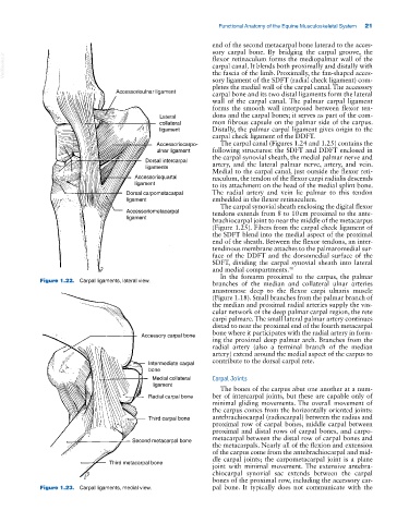
             Figure 1.22.  Carpal ligaments, lateral view.
                                                                 branches of the  median and collateral ulnar arteries
                                                                 anastomose deep to the flexor carpi ulnaris muscle
                                                                 (Figure 1.18). Small branches from the palmar branch of
                                                                 the median and proximal radial arteries supply the vas­
                                                                 cular network of the deep palmar carpal region, the rete
                                                                 carpi palmare. The small lateral palmar artery continues
                                                                 distad to near the proximal end of the fourth metacarpal
                                                                 bone where it participates with the radial artery in form­
                                           Accessory carpal bone
                                                                 ing the proximal deep palmar arch. Branches from the
                                                                 radial artery (also a terminal branch of the median
                                                                 artery) extend around the medial aspect of the carpus to
                                                                 contribute to the dorsal carpal rete.
                                             Intermediate carpal
                                             bone
                                               Medial collateral  Carpal Joints
                                               ligament
                                                                   The bones of the carpus abut one another at a num­
                                             Radial carpal bone  ber of intercarpal joints, but these are capable only of
                                                                 minimal gliding movements. The overall movement of
                                                                 the carpus comes from the horizontally oriented joints:
                                             Third carpal bone   antebrachiocarpal (radiocarpal) between the radius and
                                                                 proximal row of carpal bones, middle carpal between
                                                                 proximal and distal rows of carpal bones, and carpo­
                                                                 metacarpal between the distal row of carpal bones and
                                         Second metacarpal bone
                                                                 the metacarpals. Nearly all of the flexion and extension
                                                                 of the carpus come from the antebrachiocarpal and mid­
                                                                 dle carpal joints; the carpometacarpal joint is a plane
                                  Third metacarpal bone
                                                                 joint with minimal movement. The extensive antebra­
                                                                 chiocarpal synovial sac extends between the carpal
                                                                 bones of the proximal row, including the accessory car­
             Figure 1.23.  Carpal ligaments, medial view.        pal bone. It typically does not communicate with the
   50   51   52   53   54   55   56   57   58   59   60