
Page 633 - Adams and Stashak's Lameness in Horses, 7th Edition
P. 633
Lameness of the Proximal Limb 599
be caused by unrecognized congenital ALD that wors-
ens, trauma to the physis or the epiphysis, lameness on a
VetBooks.ir large body mass, inappropriate exercise, lack of exercise,
contralateral limb, overnutrition as a foal which induces
excess exercise, or poor conformation with growth. As
13
an example, foals that have narrow chests at birth and
yet straight limbs have a propensity to develop a wide
chest that may lead to carpal varus in the future. In these
cases it is good to examine the mare and possibly the
stallion to try to determine the type of conformation
that may influence the joints in the future. If trauma to
the physis or epiphysis is severe enough, the growth may
actually stop on that side due to damage to the physeal
cartilage, sometimes leading to bone bridging across the
physis. Any skin abrasion over a physis should be
39
thoroughly explored to be sure that contamination and
subsequent septic physitis does not develop.
Clinical Signs and Diagnosis
The most important information that a veterinarian
can give an owner about foals is that they require con-
stant monitoring during growth. Foals can be consid-
ered to be easily deformable due to a number of factors
during growth, as mentioned above, and they require at
least weekly monitoring to stay ahead of rapid changes.
For instance, a lameness of any cause in any limb may
cause secondary ALD in other limbs. Therefore, during
history taking, it is important to know how often the
foal has been monitored and the changes noted by the
owner since birth. The history should reveal whether
these foals were normal at birth, if they were premature,
the diet of the mare and foal, and when the ALD was
first noticed.
The foal should be examined both statically and
dynamically. It should be allowed to freely walk around
so as not to induce an abnormal stance through han-
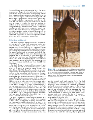
dling (Figure 5.2). This should be done for several min- Figure 5.2. A foal demonstrating normal degree of carpal valgus
utes because clinical impressions can easily change based expected after birth in the left forelimb and a moderate carpal valgus
on how the foal is standing. It is also common to exam- of the right forelimb. Notice that the foal is standing fairly square on
its own, providing a good examination. However, the examination
ine the limb with the veterinarian standing at the foal’s findings should be an assessment based on several minutes of
shoulder, looking straight down through the radius, car- examination and movement.
pus, and third metacarpus to assess ALD. In addition,
because rotational deformities are common with carpal
ALD, care must be taken not to overinterpret the sever- moving around freely and standing freely. The foal
ity. For instance, it is common to see outward rotation should also be walked to characterize foot flight. For
of the limb in addition to a carpal valgus deformity. If instance, it is not uncommon in foals with carpal varus
the foal is examined from directly in front of the foal’s to break over the dorsolateral aspect of the foot, in
head, then this rotational deformity can make the ALD which case they may actually swing the limb out. The
look worse than it really is. It is important to remember same is true with carpal valgus, in which the foal may
to stand directly in front of the face of the particular swing the leg medially due to dorsomedial break‐over.
carpus. In the case of the previous example, the exam- The conformation of the hoof can often help in deter-
iner must stand dorsolateral to the midline plane of the mining break‐over, because the site of break‐over—
foal to accurately assess each carpus. either dorsolateral or dorsomedial—will be worn
In some instances the author recommends that peri- compared with the other side.
odic photographs or videos be e‐mailed for review, and During physical examination it is important to iden-
in these cases it is important to remind the owner that tify the site of deformity and estimate its severity, char-
the photographs must be taken directly in front of the acterizing it as mild, moderate, or severe. Some people
face of the carpus. It is common to recommend taking advocate the use of a goniometer; however, classifying
several photographs starting at the very front of the foal these into mild, moderate, and severe is sometimes just
and rotating laterally right and left in front of the knee. as effective. It is important to jog these foals or let them
During physical examination, the foal can be manu- run free in a small pen to determine whether a subtle
ally “squared up” if needed; however, that must be taken lameness is present, especially in an opposing limb, and
into consideration along with assessment of the foal to palpate the limbs for effusion, physeal pain, and