
Page 634 - Adams and Stashak's Lameness in Horses, 7th Edition
P. 634
600 Chapter 5
swelling (Figure 5.3). It is also important to manipulate radiographically, since growth stops well before radio-
the limb to characterize any joint laxity. Physeal swelling graphic evidence is seen. In particular, assessment of
VetBooks.ir physitis can be insidious in onset and severity. mated with radiographs, because it is not uncommon
distal radial physeal closure cannot be accurately esti-
in particular should be palpated for pain because septic
The above physical examination techniques can only
for the physis to remain visible even after growth has
be performed if the foal is sound and standing. When the stopped.
foal is recumbent, it can be observed while trying to Both dorsopalmar and lateromedial views are neces-
stand. If this cannot be accomplished and if the owner sary. On the dorsopalmar views, lines are drawn dissect-
has not seen the foal stand and nurse, then serum IgG ing both the distal radius and the proximal third
concentrations in the foal should be assessed. If the foal metacarpal bone; the area where the lines intersect helps
is lying down, joint laxity must be assessed, and if there to describe the location within the joint in which
19
is severe ALD, it is important to assess whether this is the deviation is occurring (Figure 5.4). For instance, if
reducible and if swelling is present anywhere on the the intersection occurs within the joints, then cuboidal
limb. The limb should be put through a full range of bone malformation often is the cause of the ALD. A
motion to assess whether pain is present and to assess goniometer or digital measurement can be used on the
the full range of extension and flexion. radiograph to measure the severity of angulation. In
addition, lines can be drawn through the joint surfaces
and the distal radial physis, and the line or lines that
Diagnostic Imaging
deviate from perpendicular to the third metacarpal bone
Radiographs of the carpus should be performed to can be identified as the site of deviation.
include the distal radius and proximal cannon bone for Care must be taken not to overinterpret these results.
points of reference for measuring ALD. Some clinicians It is not uncommon to have to sedate foals to acquire
advocate using long film detectors to do this; however, radiographs, and the sedated stance may not truly repre-
appropriate assessment can usually be accomplished sent the severity of deformity. In most cases sedation can
using standard detectors. It is important to remember make any degree of joint laxity worsen the appearance
that the growth status of the physes often cannot be of the radiographic findings.
accurately assessed radiographically. For instance, In addition to measuring angles, the degree of ossifi-
characterizing a physis as open or closed is difficult cation, malformation, and damage to the joint must be
characterized. It is recommended that a full series of car-
pal radiographs be taken if initial films show any signs
of damage, because subtle damage can affect the prog-
nosis. Any foal deemed to be premature or dysmature
should have a radiographic examination of both carpi
and other joints such as tarsi since incomplete ossification
of the cuboidal bones is not uncommon. Dorsopalmar
and lateromedial views of the carpus should be obtained.
A grading system has been developed called the skeletal
ossification index (SOI), which has been correlated
1
with gestational age and body weight, but not with
prognosis for soundness.
It is not uncommon in the physes to see metaphyseal
flare, asymmetrical widening of the physes, sclerosis of
the physes (which usually occurs on the concave sur-
face), physeal widening (which usually occurs on the
convex side of the deformity), epiphyseal wedging, car-
pal bone wedging (which primarily occurs in the third
carpal bone), distal displacement of the ulnar carpal
bone or the fourth metacarpal bone, and increased angu-
lation of the head of the fourth metacarpal bone. 14,15,98
Although it is important to characterize these findings,
their significance on prognosis is questionable. In addi-
tion, any osteochondral fragments, signs of physeal
trauma, septic physitis, osteomyelitis, or arthritis must
be taken into consideration because their presence sig-
nificantly affects the prognosis. In general, most people
characterize the degree of angulation as mild (5°–10°),
moderate (15°–25°), or severe (more than 25°).
Treatment
Treatment of ALD of the carpus depends on the
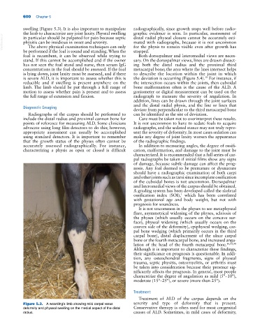
Figure 5.3. A weanling’s limb showing mild carpal varus severity and type of deformity that is present.
deformity and physeal swelling on the medial aspect of the distal Conservative therapy is often used for most congenital
radius. causes of ALD. Sometimes, in mild cases of deformity,